

Percutaneous tumor ablation with radiofrequency
- PMID: 11900230
- PMCID: PMC2408948
- DOI: 10.1002/cncr.10234
Percutaneous tumor ablation with radiofrequency
Abstract
Background: Radiofrequency thermal ablation (RFA) is a new minimally invasive treatment for localized cancer. Minimally invasive surgical options require less resources, time, recovery, and cost, and often offer reduced morbidity and mortality, compared with more invasive methods. To be useful, image-guided, minimally invasive, local treatments will have to meet those expectations without sacrificing efficacy.
Methods: Image-guided, local cancer treatment relies on the assumption that local disease control may improve survival. Recent developments in ablative techniques are being applied to patients with inoperable, small, or solitary liver tumors, recurrent metachronous hereditary renal cell carcinoma, and neoplasms in the bone, lung, breast, and adrenal gland.
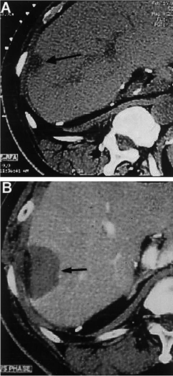
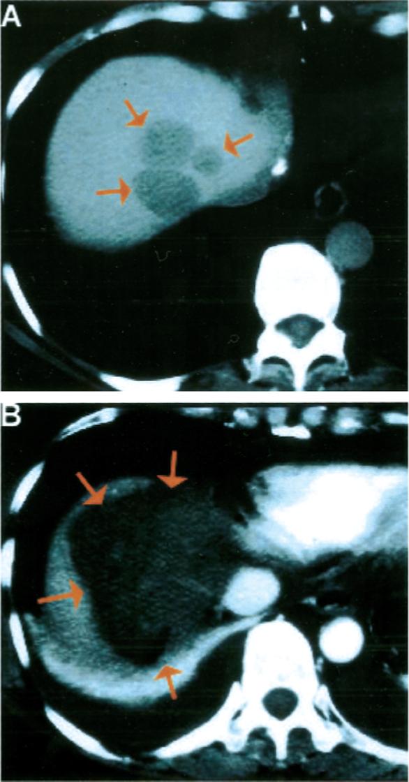
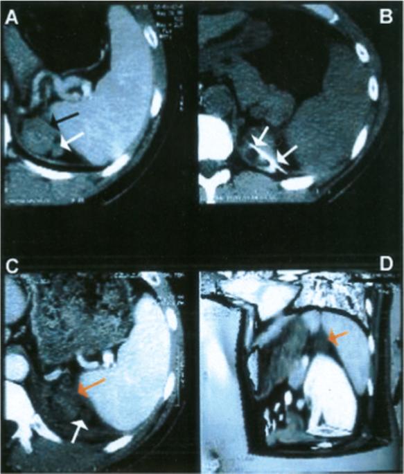
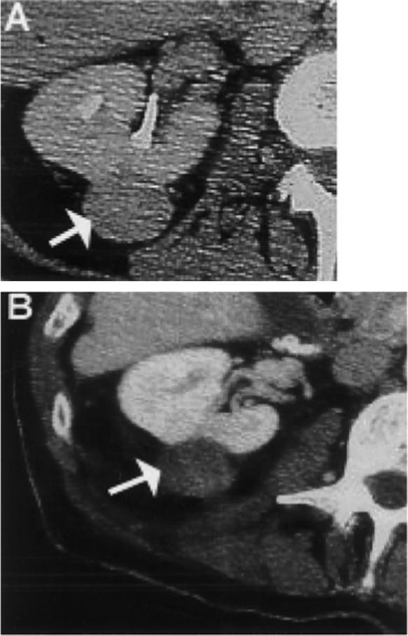
Results: Recent refinements in ablation technology enable large tumor volumes to be treated with image-guided needle placement, either percutaneously, laparoscopically, or with open surgery. Local disease control potentially could result in improved survival, or enhanced operability.
Conclusions: Consensus indications in oncology are ill-defined, despite widespread proliferation of the technology. A brief review is presented of the current status of image-guided tumor ablation therapy. More rigorous scientific review, long-term follow-up, and randomized prospective trials are needed to help define the role of RFA in oncology.
Figures
References
-
- Adams F. Genuine works of Hippocrates. William Wood and Company; New York: 1886. translator.
-
- DeSanctis JT, Goldberg SN, Mueller PR. Percutaneous treatment of hepatic neoplasms: a review of current techniques. Cardiovasc Intervent Radiol. 1998;21:273–96. - PubMed
-
- Scudamore CH, Patterson EJ, Shapiro J, Buczkowski AK. Liver tumor ablation techniques. J Invest Surg. 1997;10:157–64. - PubMed
-
- Oturai AB, Jensen K, Eriksen J, Madsen F. Neurosurgery for trigeminal neuralgia: comparison of alcohol block, neurectomy and radiofrequency coagulation. Clin J Pain. 1996;12:311–5. - PubMed
-
- Rosenthal DI, Hornicek FJ, Wolfe MW, Jennings LC, Gebhardt MC, Mankin HJ. Percutaneous radiofrequency coagulation of osteoid osteoma compared with operative treatment. J Bone Joint Surg Am. 1998;80:815–21. - PubMed
Publication types
MeSH terms
Grants and funding
LinkOut - more resources
Full Text Sources
Other Literature Sources
Medical
