. 2002 Mar 26;41(12):3991-4001.
doi: 10.1021/bi011922x.

Molecular features of the copper binding sites in the octarepeat domain of the prion protein

Affiliations

Molecular features of the copper binding sites in the octarepeat domain of the prion protein

Colin S Burns et al. Biochemistry. .

Abstract

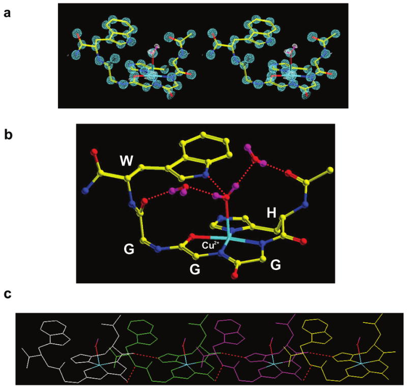
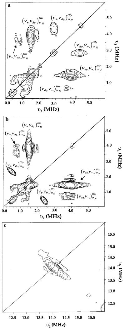
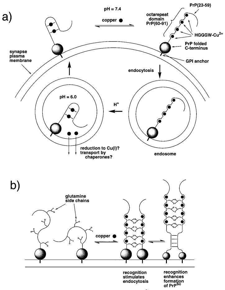
Recent evidence suggests that the prion protein (PrP) is a copper binding protein. The N-terminal region of human PrP contains four sequential copies of the highly conserved octarepeat sequence PHGGGWGQ spanning residues 60-91. This region selectively binds Cu2+ in vivo. In a previous study using peptide design, EPR, and CD spectroscopy, we showed that the HGGGW segment within each octarepeat comprises the fundamental Cu2+ binding unit [Aronoff-Spencer et al. (2000) Biochemistry 40, 13760-13771]. Here we present the first atomic resolution view of the copper binding site within an octarepeat. The crystal structure of HGGGW in a complex with Cu2+ reveals equatorial coordination by the histidine imidazole, two deprotonated glycine amides, and a glycine carbonyl, along with an axial water bridging to the Trp indole. Companion S-band EPR, X-band ESEEM, and HYSCORE experiments performed on a library of 15N-labeled peptides indicate that the structure of the copper binding site in HGGGW and PHGGGWGQ in solution is consistent with that of the crystal structure. Moreover, EPR performed on PrP(23-28, 57-91) and an 15N-labeled analogue demonstrates that the identified structure is maintained in the full PrP octarepeat domain. It has been shown that copper stimulates PrP endocytosis. The identified Gly-Cu linkage is unstable below pH approximately 6.5 and thus suggests a pH-dependent molecular mechanism by which PrP detects Cu2+ in the extracellular matrix or releases PrP-bound Cu2+ within the endosome. The structure also reveals an unusual complementary interaction between copper-structured HGGGW units that may facilitate molecular recognition between prion proteins, thereby suggesting a mechanism for transmembrane signaling and perhaps conversion to the pathogenic form.


Figures

Figure 1
Figure 1
Sequence of PrP and the octarepeat domain. (a) This sequence representation of PrPC locates the globular C-terminal domain, the glycosylphosphatidylinositol (GPI) membrane anchor, and the octarepeat domain. Also shown is a flexible region implicated in multimerization that accompanies PrPC → PrPSc conversion. Cu2+ binding within the octarepeats involves the specific residues HGGGW (underlined). (b) Alignment of the octarepeat domain from several species. Note that the only sequence variation within the HGGGW segments is either variation of the third glycine (to serine in two mouse octarepeats) or insertion of an additional glycine (in the porcine sequence).
Figure 2
Figure 2
Crystal structure (0.7 Å resolution) of the HGGGW segment in complex with Cu2+. (a) A stereo representation of the electron density contoured at 2 σ is shown in blue. A difference map (white) reveals hydrogen density for the axially bound water. (b) This molecular representation shows how copper coordination is from the histidine imidazole and deprotonated amides from the next two glycines. In addition, the NH of the indole is within hydrogen bonding distance to the oxygen of the axial water. Two additional intramolecular ordered water molecules are also shown. (c) The red dashed lines show intermolecular hydrogen bond contacts identified in the crystal. These four copies of the copper binding units suggest a possible way in which the full octarepeat domain orders.
Figure 3
Figure 3
The mI = −1/2 line from the S-band EPR (3.5 GHz) of Cu2+ bound to 15N-labeled HGGGW and PHGGGWGQ. For HGGGW, a change in multiplet structure is observed only when the first two glycines are 15N-labeled, demonstrating that these nitrogens coordinate Cu2+. The vertical lines drawn from the most prominent features of the unlabeled spectra are included to guide the eye. The bottom two spectra are simulations where sim. 1 is for three equivalent 14N (aN = 13 G) and a hydrogen (aH = 10 G) and sim. 2 has one 14N replaced with an 15N. PHGGGWGQ shows the same pattern as HGGGW, indicating that the first two glycines coordinate to the copper center.
Figure 4
Figure 4
Stimulated ESEEM spectra of HGGGW, PHGGGWGQ, and PrP(23–28, 57–91). (a) Spectra of unlabeled species with assignments, based on comparison to spectra obtained from the 15N-labeled peptides (see panel b), shown by the grids at the top. All spectra from unlabeled constructs show coupling to the remote NH of the imidazole and a noncoordinated amide nitrogen. (b) These spectra of 15N-labeled HGGGW and PrP(23–28, 57–91, 15N-64,72,80,88) show only those transitions due to the remote NH of the His imidazole consistent with the finding that the third glycine in each HGGGW is not coordinated to the Cu2+. Parameters for data acquisition were as follows: temperature, 4.2 K; π/2 pulse widths, 20 ns; repetition rate, 13 Hz; τ, 140 ns; T increment, 20 ns; 30 averages per point.
Figure 5
Figure 5
HYSCORE spectra of HGGGW with (b) and without (a) 15N-substitution at the third glycine amide. Peaks assigned to imidizole nuclear quadrupolar (NQI) and double quantum (DQ) transitions are correlated in the spectra and remain so after substitution of 15N. In spectrum b, the glycine NQI and DQ features are lost and replaced by peaks at 0.47 and 2.43 MHz corresponding to a dipolar coupling to the metal center. Panel c shows the spectra consistent with weakly coupled protons. Parameters for data acquisition were as follows: temperature, 4.2 K; π/2 pulse widths, 20 ns; repetition rate, 53 Hz; τ, 160 ns; T increment, 40 ns; 40 averages per point. Initial t1,2 = 40 ns, t1,2 step = 40 ns.
Figure 6
Figure 6
Working hypotheses for how the prion protein functions to transport Cu2+ through endocytosis and how the octarepeat domain participates in the formation of pathogenic PrPSc. (a) At extracellular pH, each octarepeat binds a single Cu2+. Within the endosome at pH ≈ 6.0, protonation of the amides competes with Cu2+ coordination, thereby lowering the affinity for the metal ion. There is no current information as to how PrP binds remaining Cu2+ at low pH; however, some experimental evidence points to multiple His coordination (see Discussion). (b) The glutamines in each octarepeat do not participate in Cu2+ coordination yet are highly conserved in mammalian prions. Copper orders the octarepeat domain at pH 7.4 bringing the glutamines into close proximity to one another. The Gln side chains facilitate intermolecular recognition between neighboring, membrane-bound PrPs. This noncovalent cross-linking may be a molecular signal that stimulates endocytosis or transmembrane signaling. When accompanied by rare events leading to partial unfolding of the globular domain, the cross-linked species allows for recognition between PrPs in the adjacent region PrP(90–120), thereby facilitating formation of PrPSc.

References

    1. Prusiner SB. Proc Natl Acad Sci USA. 1998;95:13363–13383. - PMC - PubMed
    1. Whittal RM, Ball HL, Cohen FE, Burlingame AL, Prusiner SB, Baldwin MA. Protein Sci. 2000;9:332–343. - PMC - PubMed
    1. Brown DR, Qin K, Herms JW, Madlung A, Manson J, Strome R, Fraser PE, Kruck T, von Bohlen A, Schulz-Schaeffer W, Giese A, Westaway D, Kretzschmar H. Nature. 1997;390:684–687. - PubMed
    1. Aronoff-Spencer E, Burns CS, Avdievich NI, Gerfen GJ, Peisach J, Antholine WE, Ball HL, Cohen FE, Prusiner SB, Millhauser GL. Biochemistry. 2000;39:13760–13771. - PMC - PubMed
    1. Bonomo RP, Impellizzeri G, Pappalardo G, Rizzarelli E, Tabbi G. Chem–Eur J. 2000;6:4195–4202. - PubMed

Publication types