

Sandlike appearance of Virchow-Robin spaces in early multiple sclerosis: a novel neuroradiologic marker
- PMID: 11901003
- PMCID: PMC7975312
Sandlike appearance of Virchow-Robin spaces in early multiple sclerosis: a novel neuroradiologic marker
Abstract
Background and purpose: The distinctive hyperintensity of multiple sclerosis (MS) lesions on T2-weighted brain MR images is well recognized. However, Virchow-Robin spaces (VRSs), especially in early MS, have not been described. Our purpose was to determine the frequency of VRSs in recent-onset MS.
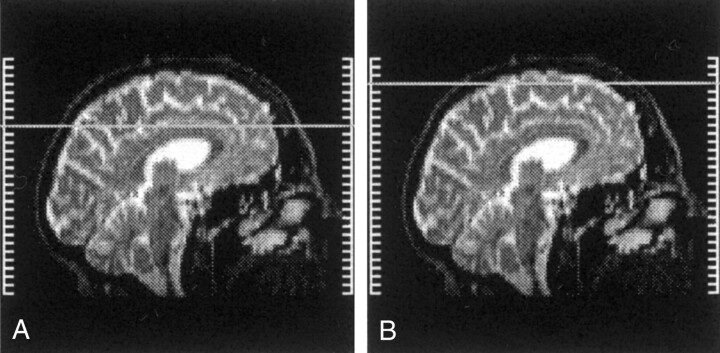
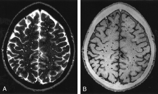
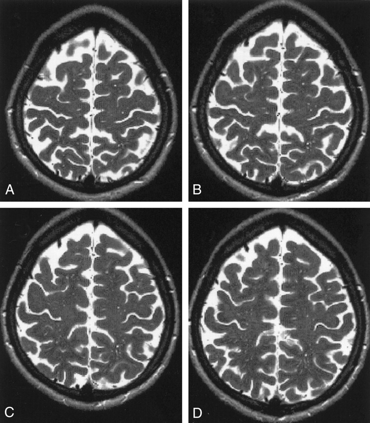
Methods: Brain MR imaging was performed in 71 patients (mean age, 26.8 years; range, 20-41 years; 47 women, 24 men) within 3 months of MS onset. Proton density-, T2-, and T1-weighted images were obtained. Age-and sex-matched control subjects (mean age, 27.2 years; range, 22-41 years; 38 women, 22 men) who underwent brain MR imaging as a part of headache evaluation, and findings that were interpreted as normal served as controls. On high-convexity images (axial sections above the upper corpus callosum border), VRSs were identified as small (<2-mm diameter) sandlike areas isointense to CSF. VRSs were graded 0-3.
Results: VRSs were visualized in high-convexity white matter in 55% of patients and 7% of control subjects (P <.001). In patients, 15% of VRSs were grade 1 (fewer than four), 23% were grade 2 (four to seven), and 62% were grade 3 (more than seven). In control subjects, all identified VRSs were grade 1. Among patients with and those without VRSs, age at onset, neurologic disability, and specific functional system involvement or mono- versus polysymptomatic involvement at onset did not differ.
Conclusion: VRSs were more frequent in patients with recent-onset MS than in control subjects. The sandlike appearance of VRSs may be a neuroradiologic marker that reflects early inflammatory changes in MS.
Figures
Comment in
-
"Dilated perivascular spaces: a hallmark of mild traumatic brain injury"--a new paradigm?AJNR Am J Neuroradiol. 2005 Apr;26(4):692-3. AJNR Am J Neuroradiol. 2005. PMID: 15814904 Free PMC article. No abstract available.
References
-
- Hutchings M, Weller RO. Anatomical relationships of the pia mater to cerebral blood vessels in man. J Neurosurg 1986;65:316–325 - PubMed
-
- Jungreis CA, Kanal E, Hirsch WL, et al. Normal perivascular spaces mimicking lacunar infarction: MR imaging. Radiology 1988;169:101–104 - PubMed
-
- Bokura H, Kobayashi S, Yamaguchi S. Distinguishing silent lacunar infarction from enlarged Virchow-Robin spaces: a magnetic resonance imaging and pathological study. J Neurol 1998;245:116–122 - PubMed
MeSH terms
LinkOut - more resources
Full Text Sources
Medical