

Case Reports
Diagnostic pitfall: atypical cerebral venous drainage via the vertebral venous system
Affiliations
- PMID: 11901009
- PMCID: PMC7975308
Item in Clipboard
Case Reports
Diagnostic pitfall: atypical cerebral venous drainage via the vertebral venous system
AJNR Am J Neuroradiol.
2002 Mar.
Abstract
We report a case of atypical cerebral venous drainage in a 38-year-old woman with symptoms of benign paroxysmal positional vertigo. Thrombosis of the left internal jugular vein and sigmoid sinus was suspected on the basis of spin-echo and time-of-flight MR findings, but multisection CT angiograms showed a patent sigmoid sinus and predominant drainage via the emissary veins toward the vertebral plexus, with only a minor contribution of the jugular veins. This case illustrates the variability of the venous anatomy in the craniocervical region.
Figures
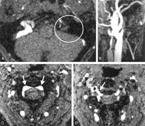
Cervical 2D TOF MR angiograms. A, Axial source image depicts no contrast enhancement within the left sinus sigmoideus (circle). B, Maximum intensity projection (MIP) reconstruction image of the left IJV depicts only a short proximal vessel segment with regular contrast enhancement. C, Axial source image obtained at the level of C2 shows prominent epidural veins of the anterior intraspinal system (arrows). D, Axial source image obtained at the level of the C2–3 intervertebral space shows that a radicular vein (straight arrow) connects the anterior intraspinal system to the right vertebral vein (curved arrow) next to the vertebral artery (flow void).
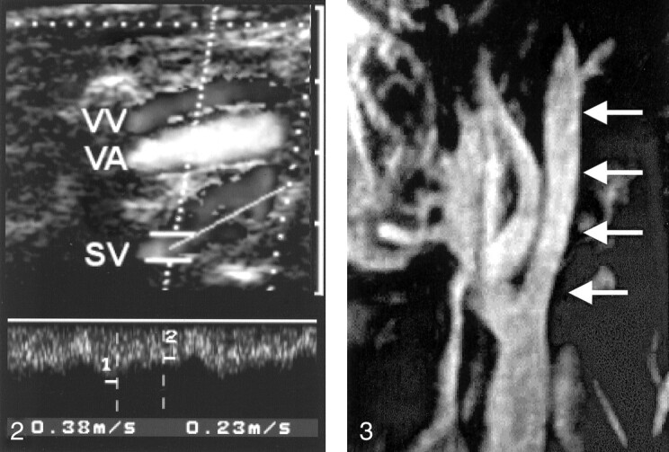
Duplex sonogram depicts the right vertebral vein (VV) and vertebral artery (VA) at the level of C4 and C5. Note the prominent segmental venous inflow (SV) with a high systolic flow velocity of 38 cm/s.
MIP of the contrast-enhanced T1-weighted gradient-echo MRV image of the left IJV. The vein has good contrast enhancement below the skull base (arrows).
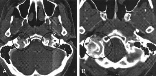
Axial images from multisection CTA. A, Bilateral asymmetrical venous contrast enhancement at the level of the jugular foramen is depicted (circles). B, Prominent posterior condylar emissary vein is depicted on the right side (circle).
References
-
- Einhäupl KM, Villringer A, Meister W, et al. Heparin treatment in sinus venous thrombosis. Lancet 1991;338:597–600 - PubMed
-
- Einhäupl K, Masuhr F. Cerebral venous and sinus thrombosis: an update. Eur J Neurol 1994;1:109–126 - PubMed
-
- Bianchi D, Maeder P, Bogousslavsky J, Schnyder P, Meuli R. Diagnosis of cerebral venous thrombosis with routine magnetic resonance: an update. Eur Neurol 1998;40:179–190 - PubMed
-
- Urchuk SN, Plewes DB. Mechanisms of flow-induced signal loss in MR angiography. J Magn Reson Imaging 1992;2:453–462 - PubMed
-
- Ozsvath RR, Casey SO, Lustrin ES, Alberico RA, Hassankhani A, Patel M. Cerebral venography: comparison of CT and MR projection venography. AJR Am J Roentgenol 1997;169:1699–1707 - PubMed
Publication types
MeSH terms
LinkOut - more resources
Full Text Sources