

Case Reports
Recognition and importance of an infraoptic anterior cerebral artery: case report
Affiliations
- PMID: 11901017
- PMCID: PMC7975300
Item in Clipboard
Case Reports
Recognition and importance of an infraoptic anterior cerebral artery: case report
AJNR Am J Neuroradiol.
2002 Mar.
Abstract
Although variations of the anterior cerebral artery (ACA)-anterior communicating artery complex are commonly identified on imaging studies, an infraoptic course of the ACA is exceedingly rare. What is believed to be the first case of an infraoptic course of the ACA discovered with MR angiography and further characterized with conventional angiography is presented. The high prevalence of associated aneurysms and the implications for surgical planning make preoperative recognition of this anomaly critical.
Figures

MR angiograms. A, Posterior oblique reformation of a 3D time-of-flight MR arteriographic data set reveals low bifurcation of the right ICA (arrowhead). Basilar tip (white arrow) and left middle cerebral artery bifurcation (black arrow) aneurysms are demonstrated. B and C, Source images from a 3D time-of-flight MR angiographic study at the level of the cavernous carotid (B) and optic chiasm (C). The right ACA (arrow) has a low origin from the ICA and ascends in a medial location between the optic nerves (arrowheads in C).
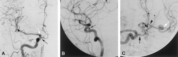
Arteriograms. A–C, Frontal (A), lateral (B), and right anterior oblique (C) views from a conventional right common carotid arteriographic study demonstrate a low origin of the right ACA (black arrow), proximal to the ophthalmic artery. A small infundibulum (white arrow in A) demarcates the origin of the ophthalmic artery. The A1 segment of the ACA has a characteristic horizontal-medial course (as it passes under the ipsilateral optic nerve) before ascending to join a normally positioned AcomA. Reflux of contrast material across the patent AcomA illustrates the normal appearance of the contralateral A1 segment (arrowhead in C). The left middle cerebral artery bifurcation aneurysm (white arrow in C) is demonstrated.
References
-
- Spinnato S, Pasqualin A, Chioffi F, Da Pian R. Infraoptic course of the anterior cerebral artery associated with an anterior communicating artery aneurysm: anatomic case report and embryological considerations. Neurosurgery 1999;44:1315–1319 - PubMed
-
- Maurer J, Maurer E, Perneczky A. Surgically verified variations in the A1 segment of the anterior cerebral artery: report of two cases. J Neurosurg 1991;75:950–953 - PubMed
-
- Fujimoto S, Murakami M. Anomalous branch of the internal carotid artery supplying circulation of the anterior cerebral artery: case report. J Neurosurg 1983;58:941–946 - PubMed
-
- Nutik S, Dilenge D. Carotid-anterior cerebral artery anastomosis: case report. J Neurosurg 1976;44:378–382 - PubMed
Publication types
MeSH terms
LinkOut - more resources
Full Text Sources