

Case Reports
MR imaging and MR spectroscopy in rhizomelic chondrodysplasia punctata
Affiliations
- PMID: 11901023
- PMCID: PMC7975319
Item in Clipboard
Case Reports
MR imaging and MR spectroscopy in rhizomelic chondrodysplasia punctata
AJNR Am J Neuroradiol.
2002 Mar.
Abstract
A case of rhizomelic chondrodysplasia punctata was investigated with MR imaging of the brain and hydrogen-1 MR spectroscopy of the brain and blood. Areas with abnormal signal hyperintensity on T2-weighted images or hypointensity on T1-weighted images were detected in the subcortical white matter. MR spectroscopy of the brain showed that normal-appearing white matter was characterized by increased levels of mobile lipids and myo-inositol, reduced levels of choline, and the presence of acetate. The importance of these metabolic anomalies is correlated to the deficiency in plasmalogen biosynthesis.
Figures

MR images. A, Axial T1-weighted image of the basal ganglia shows small delivery-related hemorrhage over the temporal convexity and bilateral areas (arrows) of low signal intensity in the white matter of the most anterior portion of the superior frontal gyrus. Hypointense areas are more pronounced on the left. Note the encysted cavum septi pellucidi. B, Coronal T2-weighted image shows bilateral areas (arrows) of high signal intensity in the superior parietal lobules. Hyperintense areas are more pronounced on the left.
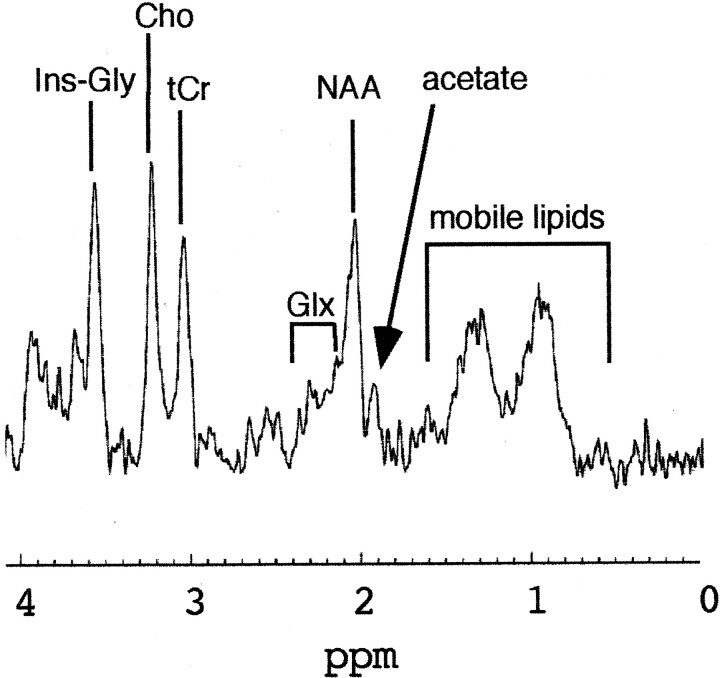
1H MR spectrum obtained at with the STEAM sequence (TE, 20 milliseconds) in the occipitoparietal white matter. The dominant feature of the spectrum is the high lipid and Ins-Gly content. The presence of an unusual resonance at 1.95 ppm was detected and assigned to acetate.
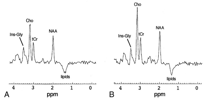
Long-TE (135 milliseconds) 1H spectra obtained at 1.5 T with the CSI sequence in the occipitoparietal region. The main feature common to both spectra is the high Ins-Gly content. A, Gray matter. B, White matter.

1H MR spectrum in blood serum analyzed at 400 MHz shows a high concentration of lactate, creatine, and glutamate, whereas ketone body levels are normal. 1 indicates lactate; 2, myo-inositol; 3, glycine; 4, glucose; 4*, mostly glucose; 5, creatinine; 6, creatine; 7, citrate; 8, glutamine; 9, glutamate; 10, acetone; 11, lysine; 12, alanine; 13, butyrate, valerate, malonate, and succinate derivatives; 14, valine; 15, leucine; 16, α-hydroxybutyrate; 17, 3-(trimethylsilyl)propionate-2,2,3,3-d4; *, traces of glycerol from ultrafiltration.
Similar articles
-
Abnormal myelin formation in rhizomelic chondrodysplasia punctata type 2 (DHAPAT-deficiency).Dev Med Child Neurol. 2000 Jul;42(7):492-5. doi: 10.1017/s0012162200000918. Dev Med Child Neurol. 2000. PMID: 10972423
-
Delayed myelination in a rhizomelic chondrodysplasia punctata case: MR spectroscopy findings.Magn Reson Imaging. 2003 Jan;21(1):77-80. doi: 10.1016/s0730-725x(02)00625-2. Magn Reson Imaging. 2003. PMID: 12620550
-
Isolated dihydroxyacetonephosphate-acyl-transferase deficiency in rhizomelic chondrodysplasia punctata: clinical presentation, metabolic and histological findings.Eur J Pediatr. 1996 Dec;155(12):1035-9. doi: 10.1007/BF02532526. Eur J Pediatr. 1996. PMID: 8956940
-
Rhizomelic chondrodysplasia punctata: report of a case with review of the literature and correlation with other peroxisomal disorders.Pediatr Pathol Lab Med. 1995 May-Jun;15(3):503-13. doi: 10.3109/15513819509026986. Pediatr Pathol Lab Med. 1995. PMID: 8597837 Review.
-
Plasmalogens and fatty alcohols in rhizomelic chondrodysplasia punctata and Sjögren-Larsson syndrome.J Inherit Metab Dis. 2015 Jan;38(1):111-21. doi: 10.1007/s10545-014-9795-3. Epub 2014 Nov 29. J Inherit Metab Dis. 2015. PMID: 25432520 Review.
Cited by
-
Inflammatory multiple-sclerosis plaques generate characteristic metabolic profiles in cerebrospinal fluid.PLoS One. 2007 Jul 4;2(7):e595. doi: 10.1371/journal.pone.0000595. PLoS One. 2007. PMID: 17611627 Free PMC article.
-
A case of Sjögren-Larsson syndrome with minimal MR imaging findings facilitated by proton spectroscopy.Pediatr Radiol. 2012 Mar;42(3):380-2. doi: 10.1007/s00247-011-2156-6. Epub 2011 Jun 29. Pediatr Radiol. 2012. PMID: 21713441
-
Peroxisome biogenesis disorders.Transl Sci Rare Dis. 2016 Nov 7;1(2):111-144. doi: 10.3233/TRD-160003. Transl Sci Rare Dis. 2016. PMID: 29152457 Free PMC article. No abstract available.
-
From peroxisomal disorders to common neurodegenerative diseases - the role of ether phospholipids in the nervous system.FEBS Lett. 2017 Sep;591(18):2761-2788. doi: 10.1002/1873-3468.12788. Epub 2017 Sep 7. FEBS Lett. 2017. PMID: 28796901 Free PMC article. Review.
-
Peroxisomes in brain development and function.Biochim Biophys Acta. 2016 May;1863(5):934-55. doi: 10.1016/j.bbamcr.2015.12.005. Epub 2015 Dec 11. Biochim Biophys Acta. 2016. PMID: 26686055 Free PMC article. Review.
References
-
- Wanders RJA. Peroxisomal disorders: clinical, biochemical and molecular aspects. Neurochem Res 1999;24:565–580 - PubMed
-
- Bruhn H, Kruse B, Korenke GC, et al. Proton NMR spectroscopy of cerebral metabolic alterations in infantile peroxisomal disorders. J Comput Assist Tomogr 1992;16:335–344 - PubMed
-
- Sztriha L, Al-Gazali LI, Wanders RJ, Ofman R, Nork M, Lestringant GG. Abnormal myelin formation in rhizomelic chondrodysplasia punctata type 2 (DHAPAT-deficiency). Dev Med Child Neurol 2000;42:492–495 - PubMed
Publication types
MeSH terms
Substances
LinkOut - more resources
Full Text Sources