. 2002 Mar;109(6):745-53.
doi: 10.1172/JCI7272.

Protective immunity against atherosclerosis carried by B cells of hypercholesterolemic mice

Affiliations

Protective immunity against atherosclerosis carried by B cells of hypercholesterolemic mice

Giuseppina Caligiuri et al. J Clin Invest. 2002 Mar.

Abstract

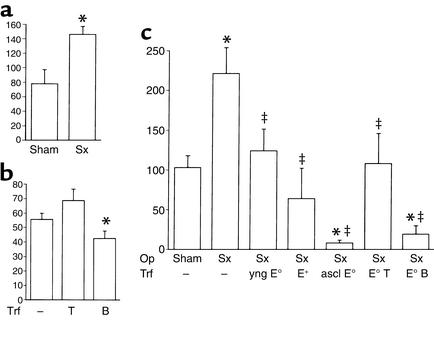
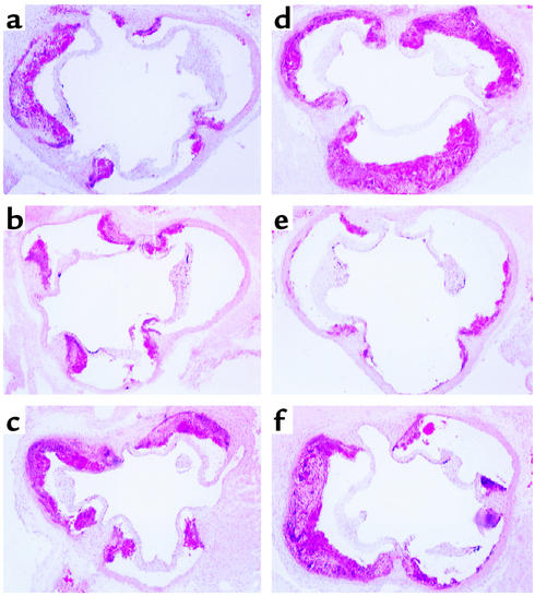
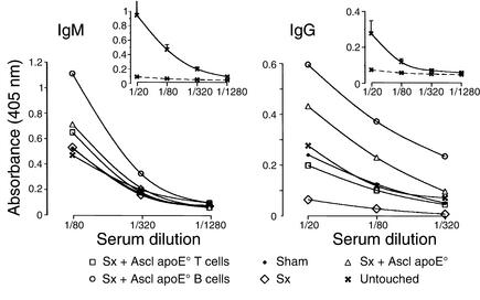
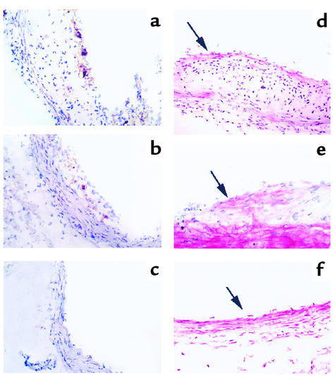
Atherosclerosis is characterized by vascular inflammation and associated with systemic and local immune responses to oxidized LDL (oxLDL) and other antigens. Since immunization with oxLDL reduces atherosclerosis, we hypothesized that the disease might be associated with development of protective immunity. Here we show that spleen-associated immune activity protects against atherosclerosis. Splenectomy dramatically aggravated atherosclerosis in hypercholesterolemic apoE knockout (apoE degrees ) mice. Transfer of spleen cells from atherosclerotic apoE degrees mice significantly reduced disease development in young apoE degrees mice. To identify the protective subset, donor spleen cells were divided into B and T cells by immunomagnetic separation before transfer. Protection was conferred by B cells, which reduced disease in splenectomized apoE degrees mice to one-fourth of that in splenectomized apoE degrees controls. Protection could also be demonstrated in intact, nonsplenectomized mice and was associated with an increase in antibody titers to oxLDL. Fewer CD4(+) T cells were found in lesions of protected mice, suggesting a role for T-B cell cooperation. These results demonstrate that B cell-associated protective immunity develops during atherosclerosis and reduces disease progression.


Figures

Figure 1
Figure 1
Effect of splenectomy and cell transfer on atherosclerotic lesions in apoE° mice. Morphometric analysis of lesion size (cross-section area at the aortic root occupied by lesion, ×103μm2, mean ± SEM). Data are from a representative experiment, and additional data summarizing all experiments are displayed in Table 1. (a) Splenectomy (Sx) increases lesion formation. Lesion size 12 weeks after Sx was significantly increased (P < 0.05, n = 6 per group) when compared with sham-operated apoE° mice. (b) Transfer (Trf) of B cells but not T cells from atherosclerotic apoE° mice reduces lesion formation in sham-operated apoE° mice. Lesions in the latter (n = 6) were significantly larger than those in B cell–transferred mice (P < 0.05, n = 7) but not different from those in T cell–transferred ones (n = 5). (c) Spleen cell transfer rescues apoE° mice from the proatherogenic effect of splenectomy. Average lesion sizes are shown for sham-operated (n = 8) and Sx (n = 7) apoE° mice, and for Sx apoE° mice who received spleen cell transfer from young apoE° mice (yng E°, n = 4 recipients), wild-type C57BL/6J mice (E+, n = 4), or atherosclerotic apoE° mice (ascl E°, n = 6) or purified spleen T cells (E° T, n = 4 recipients) or B cells (E° B, n = 4) from atherosclerotic apoE° mice. *Significantly different from sham, P < 0.05; significantly different from Sx, P < 0.05. Op, operation.
Figure 2
Figure 2
Effect of splenectomy and cell transfer on atherosclerotic lesions in the aortic root of apoE° mice. Hematoxylin/oil red O–stained lesions from the level of the aortic cusps, ×50. (a) Sham-operated mouse. (b) Sham-operated mouse that received B cells from an atherosclerotic mouse. (c) Sham-operated mouse that received T cells from an atherosclerotic mouse. (d) Splenectomized mouse. (e) Splenectomized mouse that received B cells from an atherosclerotic mouse. (f) Splenectomized mouse that received T cells from an atherosclerotic mouse.
Figure 3
Figure 3
Effect of splenectomy and cell transfer on antibody titers to MDA-LDL. ELISA of IgM and IgG antibodies to MDA-LDL was performed in splenectomized (Sx), sham-operated, and untouched apoE° mice and in Sx mice that had received total spleen cells or isolated T or B cells from atherosclerotic, 20-week-old apoE° mice. Data show binding of IgM and IgG from diluted mouse sera to MDA-LDL–coated plates (mean absorbance). Insets: ELISA of IgM and IgG antibodies to MDA-LDL (continuous lines) and nLDL (dashed lines) in untouched apoE° mice (means ± SEM). Incubation on plates coated with native LDL resulted in absorbance levels <0.1 in all treatment groups (data not shown). n = 4 mice per group.
Figure 4
Figure 4
Long-term survival of transferred lymphocytes. (a) Kinetics of PCR reactions for GAPDH and TK. The numbers of cycles chosen for semiquantitative analysis are indicated. (b) Spleen cells of mice expressing TK under the CD4 promoter were injected into sham-operated (black bars) or splenectomized (white bars) recipients. Four weeks later, homing of transferred cells was assessed by PCR analysis of the TK gene in DNA extracts (reflecting infiltrating donor cells) and RT-PCR analysis of TK mRNA in RNA extracts (reflecting infiltrating T cells) of different organs. TK RT-PCR data were normalized to GAPDH mRNA levels determined by RT-PCR (mean ± SEM, n = 4 mice per group). M, mesenteric; P+I, para-aortic and inguinal; LN, lymph nodes.
Figure 5
Figure 5
Transfer of B cells reduces accumulation of CD4+ T cells and αSM-actin+ smooth muscle cells. (ac) Immunoperoxidase staining of CD4+ T cells in aortic lesions of (a) splenectomized apoE° mice, (b) splenectomized apoE° mice reconstituted with CD4+ T cells from atherosclerotic apoE° mice, and (c) splenectomized apoE° mice reconstituted with B cells from atherosclerotic apoE° mice (×100 magnification). Significant infiltrates of CD4+ cells are present in a and b but not in c. (df) Immunophosphatase staining of αSM-actin in aortic lesions of (d) splenectomized apoE° mice (×100), (e) splenectomized apoE° mice reconstituted with CD4+ T cells from atherosclerotic apoE° mice (×200), and (f) splenectomized apoE° mice reconstituted with B cells from atherosclerotic apoE° mice (×100). In (d), an αSM-actin+ cap is present in the lesion (arrow), and the media have reduced αSM-actin staining (compare with e and f). In e, the arrow points to αSM-actin+ cells in the lesion. In f, only a few intimal cells are discerned (arrow) and there is strong medial αSM-actin staining. (g) Quantification of CD4+ T cells. Increased infiltration of CD4+ T cells was found in lesions of splenectomized mice, while animals protected by adoptive transfer had fewer infiltrating CD4+ cells. Data are expressed as percent of all hematoxylin-stained cells in the lesions. n = 8 (sham), 7 (Sx), 4 (yng E°), 4 (E+), 6 (ascl E°), 4 (E° T), 4 (E° B). *P < 0.05 vs. sham.

Comment in

References

    1. Ross R. Atherosclerosis: an inflammatory disease. N Engl J Med. 1999;340:115–126. - PubMed
    1. Jonasson L, Holm J, Skalli O, Bondjers G, Hansson GK. Regional accumulations of T cells, macrophages, and smooth muscle cells in the human atherosclerotic plaque. Arteriosclerosis. 1986;6:131–138. - PubMed
    1. Stemme S, et al. T lymphocytes from human atherosclerotic plaques recognize oxidized LDL. Proc Natl Acad Sci USA. 1995;92:3893–3897. - PMC - PubMed
    1. Ylä-Herttuala S, et al. Rabbit and human atherosclerotic lesions contain IgG that recognizes epitopes of oxidized LDL. Arterioscler Thromb. 1994;14:32–40. - PubMed
    1. Salonen JT, et al. Autoantibody against oxidised LDL and progression of carotid atherosclerosis. Lancet. 1992;339:883–887. - PubMed

Publication types