. 2002 Mar;109(6):765-75.
doi: 10.1172/JCI14001.

The alpha(1D)-adrenergic receptor directly regulates arterial blood pressure via vasoconstriction

Affiliations

The alpha(1D)-adrenergic receptor directly regulates arterial blood pressure via vasoconstriction

Akito Tanoue et al. J Clin Invest. 2002 Mar.

Abstract

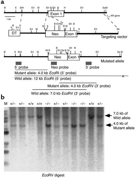
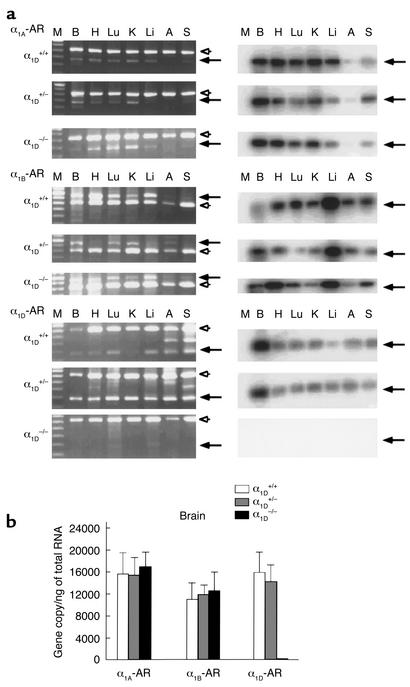
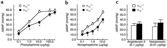
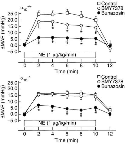
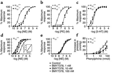
To investigate the physiological role of the alpha(1D)-adrenergic receptor (alpha(1D)-AR) subtype, we created mice lacking the alpha(1D)-AR (alpha(1D)(-/-)) by gene targeting and characterized their cardiovascular function. In alpha(1D)-/- mice, the RT-PCR did not detect any transcript of the alpha(1D)-AR in any tissue examined, and there was no apparent upregulation of other alpha(1)-AR subtypes. Radioligand binding studies showed that alpha(1)-AR binding capacity in the aorta was lost, while that in the heart was unaltered in alpha(1D)-/- mice. Non-anesthetized alpha(1D)-/- mice maintained significantly lower basal systolic and mean arterial blood pressure conditions, relative to wild-type mice, and they showed no significant change in heart rate or in cardiac function, as assessed by echocardiogram. Besides hypotension, the pressor responses to phenylephrine and norepinephrine were decreased by 30-40% in alpha(1D)-/- mice. Furthermore, the contractile response of the aorta and the pressor response of isolated perfused mesenteric arterial beds to alpha(1)-AR stimulation were markedly reduced in alpha(1D)-/- mice. We conclude that the alpha(1D)-AR participates directly in sympathetic regulation of systemic blood pressure by vasoconstriction.


Figures

Figure 1
Figure 1
Generation of α1D-AR–deficient mice. (a) Simplified restriction map around exon 1 of the α1D-AR gene and structure of the targeting vector. The coding region of the exon is boxed. Neo, PGK-neo cassette; DT, diphtheria toxin-A fragment gene; B, BamHI; E, EcoRI; EV, EcoRV; H, HindIII; Sa, SalI; SI, SacI; SII, SacII. (b) Southern blot analysis of tail DNA. DNA was digested with EcoRV, and the blot was hybridized with the 3′ probe shown in a. The 7-kb band is derived from the wild-type allele (wild) and the 4-kb band from the targeted allele (mutant).
Figure 2
Figure 2
RT-PCR analysis of the RNA from tissues of α1D+/+, α1D+/–, and α1D–/–. (a) Ethidium bromide staining of RT-PCR fragments (left). The α1A-, α1B-, and α1D-AR mRNA transcripts were detected and are shown in the upper, middle, and lower panels as 275-, 752-, and 282-bp fragments, respectively, indicated by the arrows. RT-PCR analysis was controlled by detection of the 662-bp fragment of GAPDH message, indicated by the arrowhead. Southern blots of the RT-PCR fragments are shown on the right. The specificity of the amplified fragments was assessed using 32P-labeled probes specific for each receptor subtype. M, 100-bp DNA marker; B, Brain; H, Heart; Lu, Lung; K, Kidney; Li, Liver; A, Aorta; S, Spleen. (b) TaqMan assay. Total RNA was isolated from whole brain and reverse-transcribed. Relative RNA levels of each α1-AR subtype, standardized against GAPDH levels, were obtained by semiquantitative PCR using the TaqMan system. Values represent the mean ± SEM of five independent experiments.
Figure 3
Figure 3
Blood pressure responses in α1D+/+ and α1D–/– mice. Phenylephrine (a), norepinephrine (b), angiotensin II or vasopressin (c) was injected intravenously as a bolus to male nonanesthetized α1D+/+ (open circles, n = 8) or α1D–/– mice (filled circles, n = 12) (12–18 weeks old). The effects on blood pressure are shown and expressed as the change in MAP (in mmHg). Responses to phenylephrine or norepinephrine in α1D–/– mice were significantly decreased at doses as indicated compared with the wild-type response. The maximal increase in blood pressure is shown. Points represent the mean ± SEM. *P < 0.05 as compared with α1D+/+ mice.
Figure 4
Figure 4
Effects of BMY7378 or bunazosin on blood pressure responses in α1D+/+ and α1D–/– mice under anesthesia. Inhibitory effects of BMY7378 or bunazosin on the pressor response to norepinephrine in α1D+/+ (upper) and α1D–/– (lower) mice. β-Blocker, propranolol (1 mg/kg) was preadministered, and BMY7378 (100 μg/kg) or bunazosin (10 μg/kg) was injected into male α1D+/+ or α1D–/– mice (10–12 weeks old) 10 minutes prior to continuous infusion of norepinephrine (1 μg/kg/min, for 10 minutes). Points represent the mean ± SEM of eight mice. Open squares, norepinephrine infusion; open circles, norepinephrine infusion + BMY7378 pretreatment; filled circles, norepinephrine infusion + bunazosin pretreatment. *P < 0.05 as compared with norepinephrine infusion.
Figure 5
Figure 5
Vascular contraction in α1D+/+ and α1D–/– mice. The contractile response to norepinephrine (NE), phenylephrine (PE), and serotonin (5-HT). Concentration-response curves for norepinephrine-induced (a), phenylephrine-induced (b), and serotonin-induced contractions (c) in aortic segments from α1D–/– (filled circles) or α1D+/+ mice (open circles). The results are the mean ± SEM of 15–27 preparations for NE, PE, or 5-HT. Effects of BMY7378 on norepinephrine-induced contractions in aortic segments of α1D+/+ (d) or α1D–/– mice (e). Aortic segments were exposed to vehicle (filled circles, control) or different concentrations of BMY7378 (open circle, 1 nM; filled squares, 10 nM; open squares, 100 nM), prior to the addition of cumulative concentrations of norepinephrine (NE). Inset (d): A Schild plot derived from the data from α1D+/+ mice was fitted by a straight line (R2 = 0.92) with a slope of 1.06 ± 0.11. Data represent the mean ± SEM of six different aortic segments for each group. (f) Concentration-response curves for phenylephrine-induced pressor response in perfused mesenteric arterial beds of α1D–/– mice (filled circles, n = 9) or α1D+/+ (open circles, n = 7). Two-way ANOVA showed that concentration-response curve for phenylephrine-induced pressor response of α1D–/– mice was significantly (P < 0.05) different from that of the α1D+/+ mice. *P < 0.05 as compared with α1D+/+.

References

    1. Hoffman, B.B., and Lefkowitz, R.J. 1996. Catecholamines, sympathomimetic drugs, and adrenergic receptor antagonists. In The pharmacological basis of the therapeutics. J.G. Hardman, E. Limbird, L.P.B. Molinoff, and R.W. Ruddon, editors. McGraw-Hill. New York, New York, USA. 199–248.
    1. Cotecchia S, et al. Molecular cloning and expression of the cDNA for the hamster α1-adrenergic receptor. Proc Natl Acad Sci USA. 1988;85:7159–7163. - PMC - PubMed
    1. Schwinn DA, et al. Molecular cloning and expression of the cDNA for a novel α1-adrenergic receptor subtype. J Biol Chem. 1990;265:8183–8189. - PubMed
    1. Perez DM, Piascik MT, Graham RM. Solution-phase library screening for the identification of rare clones: isolation of an α1D-adrenergic receptor cDNA. Mol Pharmacol. 1991;40:876–883. - PubMed
    1. Hirasawa A, et al. Cloning, functional expression and tissue distribution of human cDNA for the α1C-adrenergic receptor. Biochem Biophys Res Commun. 1993;195:902–909. - PubMed

Publication types

MeSH terms