. 2002 Mar;109(6):817-26.
doi: 10.1172/JCI14442.

Oxidation of the zinc-thiolate complex and uncoupling of endothelial nitric oxide synthase by peroxynitrite

Affiliations

Oxidation of the zinc-thiolate complex and uncoupling of endothelial nitric oxide synthase by peroxynitrite

Ming-Hui Zou et al. J Clin Invest. 2002 Mar.

Abstract

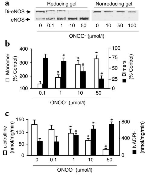
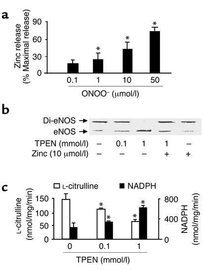
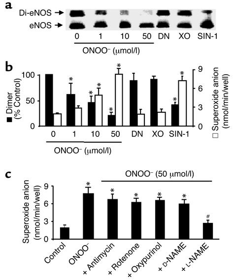
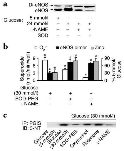
Nitric oxide (NO) is produced by NO synthase (NOS) in many cells and plays important roles in the neuronal, muscular, cardiovascular, and immune systems. In various disease conditions, all three types of NOS (neuronal, inducible, and endothelial) are reported to generate oxidants through unknown mechanisms. We present here the first evidence that peroxynitrite (ONOO(-)) releases zinc from the zinc-thiolate cluster of endothelial NOS (eNOS) and presumably forms disulfide bonds between the monomers. As a result, disruption of the otherwise SDS-resistant eNOS dimers occurs under reducing conditions. eNOS catalytic activity is exquisitely sensitive to ONOO(-), which decreases NO synthesis and increases superoxide anion (O(2)(.-)) production by the enzyme. The reducing cofactor tetrahydrobiopterin is not oxidized, nor does it prevent oxidation of eNOS by the same low concentrations of OONO(-). Furthermore, eNOS derived from endothelial cells exposed to elevated glucose produces more O(2)(.-), and, like eNOS purified from diabetic LDL receptor-deficient mice, contains less zinc and fewer SDS-resistant dimers. Hence, eNOS exposure to oxidants including ONOO(-) causes increased enzymatic uncoupling and generation of O(2)(.-) in diabetes, contributing further to endothelial cell oxidant stress. Regulation of the zinc-thiolate center of NOS by ONOO(-) provides a novel mechanism for modulation of the enzyme function in disease.


Figures

Figure 1
Figure 1
ONOO dissociates SDS-resistant dimers and alters recombinant eNOS activity. Purified eNOS was treated with ONOO (0–100 μmol/l), decomposed ONOO, or NaOH vehicle (100 mmol/l) as described in Methods. Five minutes after treatment, eNOS dimers and monomers were separated by low-temperature SDS-PAGE (6%) under reducing or nonreducing conditions. The proteins were visualized by Coomassie staining. (a) Representative blots of eNOS dimers and monomers in reducing (left) or nonreducing gels (right). (b) ONOO dissociated eNOS dimers into monomers under reducing conditions. The intensity (area times density) of dimers and monomers was determined by densitometry as described in Methods. The results were expressed as percent change compared with untreated enzyme (n = 10, *P < 0.05). (c) Inhibition by ONOO (0.1–50 μmol/l) of the rate of eNOS-dependent L-citrulline formation (n = 12) was associated with an increase in NADPH oxidation (n = 14). L-citrulline formation and NADPH oxidation by purified recombinant eNOS were each assayed as described in Methods. *P < 0.01. Di-eNOS, eNOS dimer; eNOS, eNOS monomer.
Figure 2
Figure 2
Monomer formation and activity of recombinant eNOS associated with zinc release. (a) ONOO (0.1 – 50 μmol/l) stimulated zinc release from purified eNOS (n = 9, *P < 0.01). Zinc was assayed as described in Methods and was expressed as percentage of maximal zinc release from eNOS diluted in 7 mol/l guanidine HCl. (b) Dissociation of eNOS dimers by zinc chelator TPEN. Purified eNOS was exposed to TPEN (0.1 or 1 mmol/l) or methanol (control) in the presence or absence of exogenous zinc at room temperature for 30 minutes in 0.1 M HEPES buffer, pH 7.5. eNOS protein was subjected to low-temperature SDS-PAGE under reducing conditions, and the protein was visualized by Coomassie staining. Blot shown is representative of four independent experiments. (c) The effect of TPEN on eNOS function. The activity of eNOS treated with TPEN was assayed as described in Methods. The figure represents results obtained in six independent assays (n = 6, *P < 0.05).
Figure 3
Figure 3
Comparison of ONOO reactivity with the zinc-thiolate cluster of recombinant eNOS and its cofactor, BH4. (a) BH4 is oxidized by ONOO but not by tetranitromethane (TNM). Note that 50 μmol/l ONOO, which nearly completely dissociated eNOS dimers in low-temperature SDS-PAGE under reducing conditions, did not cause detectable oxidation of BH4. These results represent 15 assays in three independent experiments. (b) Effects of BH4 and/or L-arginine on ONOO-induced dissociation of eNOS. BH4 (100 μmmol/l) and L-arginine (1 mmol/l) were added to purified eNOS 10 minutes before addition of ONOO. Blot shown is representative of three independent experiments. (c) Representative blot of three independent experiments showing that TNM, which did not oxidize BH4, results in dissociation of eNOS dimer into monomer under reducing conditions. Purified eNOS was exposed to TNM (0.1 or 1 mmol/l) or vehicle containing DMSO at room temperature for 30 minutes. eNOS dimer and monomer were assayed by low-temperature SDS-PAGE and visualized by Coomassie staining.
Figure 4
Figure 4
ONOO dissociates eNOS dimers and triggers O2.– release in cultured BAECs. (a) ONOO (1–50 μmol/l) increased dissociation of eNOS dimers in intact BAECs. The cells were treated with ONOO as described in Methods, and eNOS dimers and monomers were separated by low-temperature SDS-PAGE under reducing conditions. The eNOS protein was blotted onto nitrocellulose membranes and detected with a monoclonal antibody as described in Methods. Blot shown is representative of six independent experiments. (b) ONOO and SIN-1 decreased eNOS dimer but increased O2 release in intact BAECs. Confluent BAECs were treated with ONOO (50 mmol/I), SIN-1 (5 mmol/l), DEA-NONOate (DN; 5 mmol/l), or O2.– generated from 10 mU/ml xanthine oxidase in the presence of 0.1 mmol/l hypoxanthine (XO). The cells were rinsed twice with 2 ml PBS buffer (pH 7.4) and then exposed to calcium ionophore A23187 (10 μmol/l) for 2 hours. The amount of A23187-stimulated O2.– was measured by the SOD-inhibitable cytochrome c reduction assay as described in Methods (n = 6 or 8, *P < 0.05). (c) eNOS-dependent O2.– release in BAECs treated with ONOO. Antimycin (10 μmol/l), rotenone (10 μmol/l), oxypurinol (10 μmol/l), D-NAME (1 mmol/l), or L-NAME (1 mmol/l) was added 10 minutes after treating BAECs with ONOO and 10 minutes before the addition of A23187 (n = 10; *P < 0.05 compared with untreated cells). The increase in O2.– measured in cells exposed to ONOO was prevented by L-NAME (1 mmol/l; n = 8, #P < 0.01).
Figure 5
Figure 5
Increased monomers and decreased zinc content in eNOS derived from BAECs exposed to high glucose are associated with increased O2.– and ONOO. (a) Increased eNOS monomers in cells exposed to high glucose. Confluent BAECs were exposed to control (5 mmol/l D-glucose) or high glucose (30 mmol/l) for 3 days. The eNOS protein was separated by low-temperature SDS-PAGE under reducing conditions, blotted, and detected with a monoclonal antibody. Blot shown is representative of results from eight independent experiments. (b) Increased eNOS monomer and correlated O2.– release are associated with decreased zinc content in eNOS from cells exposed to high glucose (n = 10, #P < 0.05). The zinc content of eNOS from cells incubated in elevated glucose was assayed as described in Methods and expressed as a percentage of that in control cells exposed to 5 mmol/l glucose. Of note, both L-NAME (1 mmol/l) and SOD-PEG (500 U/ml) prevented high glucose–induced dissociation of eNOS dimers (n = 8, *P < 0.01), zinc loss (n = 5, *P < 0.01), and O2.– release (n = 6, #P < 0.05 compared with cells exposed to high glucose alone). (c) eNOS-dependent tyrosine nitration of prostacyclin synthase (PGIS) in cells exposed to high glucose. PGIS was immunoprecipitated with a polyclonal antibody against PGIS (4 μg/ml). The nitrated PGIS was detected with a monoclonal antibody against 3-nitrotyrosine (3-NT) (1:1,000; Upstate Biotechnology Inc.). Of note, SOD-PEG (500 U/ml) or L-NAME (1 mmol/l), but not oxypurinol (10 μmol/l) or rotenone (10 μmol/l), prevented the nitration of PGIS. IP, immunoprecipitation; IB, immunoblot; PGIS, prostacyclin synthase. Blot shown is representative of three independent experiments.
Figure 6
Figure 6
Increased eNOS monomer and decreased eNOS dimer and zinc content are associated with decreased eNOS activity in tissues of diabetic LDLR-KO mice. (a) Representative Western blot of eNOS protein in tissues from control and diabetic LDLR-KO mice. Tissue homogenate proteins were separated by normal-temperature SDS-PAGE under reducing conditions, blotted, and stained with a polyclonal antibody against eNOS. Blot shown is representative of three independent experiments. (b) Increased eNOS monomers are accompanied by a decrease in eNOS dimer in the tissues obtained from diabetic LDLR-KO mice. The eNOS protein in control or diabetic tissue homogenates was purified, separated by low-temperature SDS-PAGE under reducing conditions (without boiling), and detected by Coomassie staining. Results shown represent four independent experiments. (c) Decreased zinc content is associated with inhibition of NO synthesis and decreased eNOS dimer in diabetic LDLR-KO mice. The zinc content of eNOS from diabetic LDLR-KO mouse tissues was expressed as a percentage of that in normoglycemic control LDLR-KO animals. L-citrulline formation was determined in homogenates from control and diabetic animals and expressed as a percentage of that in tissues from control animals (n = 7, *P < 0.05). The assay was performed as described in Methods, and the rates of L-citrulline formation were determined to be 47 ± 11, 56 ± 19, and 43 ± 16 pmol/mg/min in liver, heart, and kidneys, respectively, of normoglycemic control LDLR-KO animals.

References

    1. Marletta MA. Nitric oxide synthase. Structure and mechanism. J Biol Chem. 1993;268:12231–12234. - PubMed
    1. Michel T, Feron O. Nitric oxide synthases: which, where, how, and why? J Clin Invest. 1997;100:2146–2152. - PMC - PubMed
    1. Nathan C, Xie QW. Regulation of biosynthesis of nitric oxide. J Biol Chem. 1994;269:13725–13728. - PubMed
    1. Raman CS, et al. Crystal structure of constitutive endothelial nitric oxide synthase: a paradigm for pterin function involving a novel metal center. Cell. 1998;95:939–950. - PubMed
    1. Ludwig ML, Marletta MA. A new decoration for nitric oxide synthase - a Zn(Cys)4 site. Structure Fold Des. 1999;7:R73–R79. - PubMed

Publication types

MeSH terms