. 2002 Mar 18;195(6):781-8.
doi: 10.1084/jem.20011714.

Pharmacological stimulation of the cholinergic antiinflammatory pathway

Affiliations

Pharmacological stimulation of the cholinergic antiinflammatory pathway

Thomas R Bernik et al. J Exp Med. .

Abstract

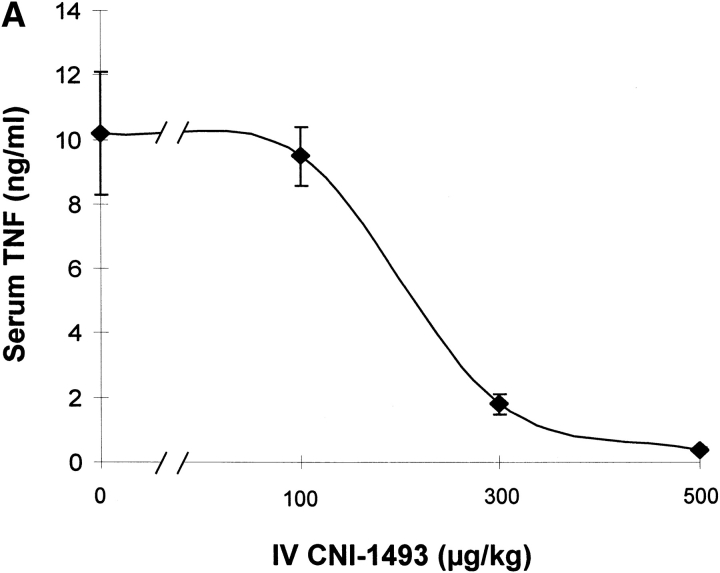
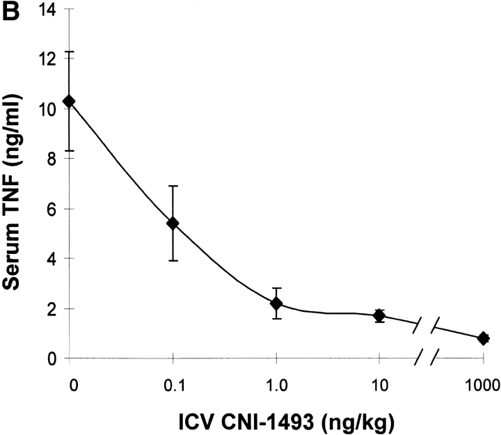
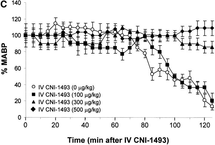
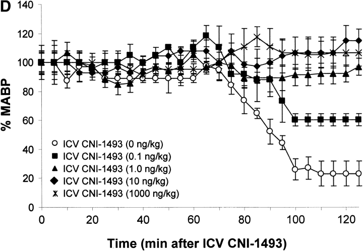
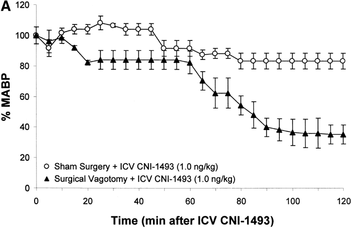
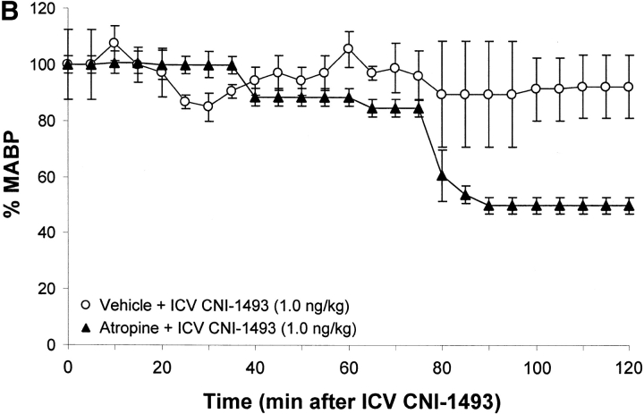
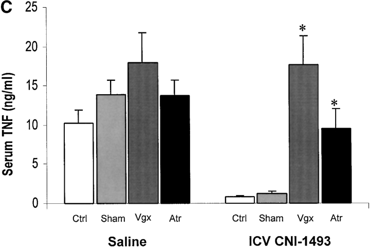
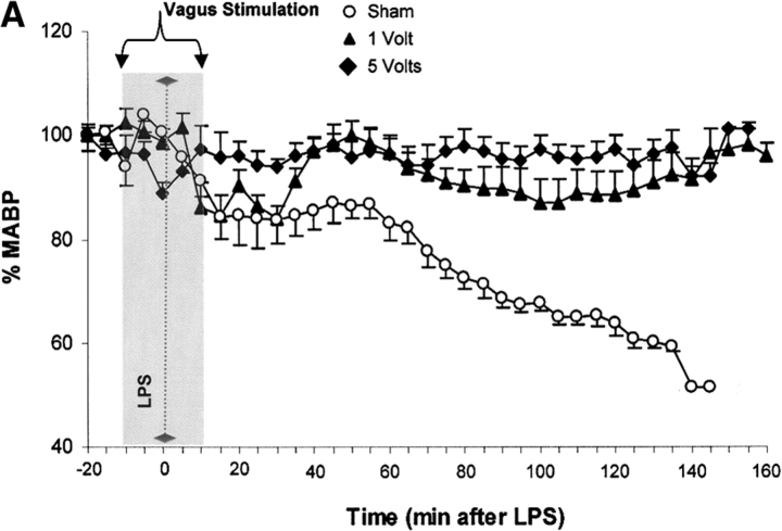
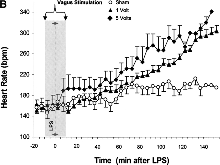
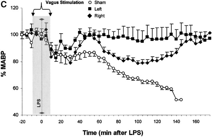
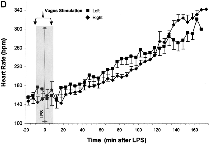
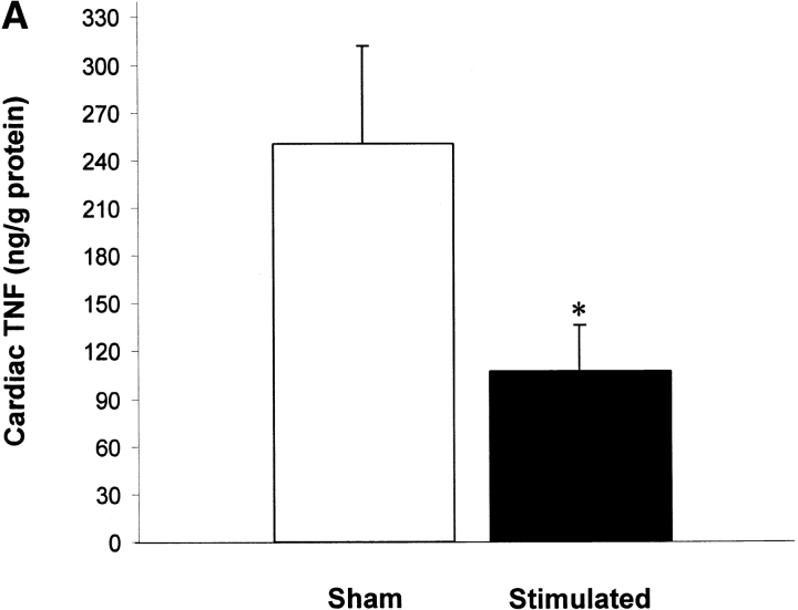
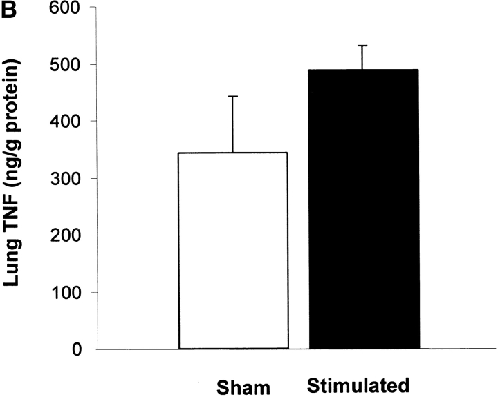
Efferent activity in the vagus nerve can prevent endotoxin-induced shock by attenuating tumor necrosis factor (TNF) synthesis. Termed the "cholinergic antiinflammatory pathway," inhibition of TNF synthesis is dependent on nicotinic alpha-bungarotoxin-sensitive acetylcholine receptors on macrophages. Vagus nerve firing is also stimulated by CNI-1493, a tetravalent guanylhydrazone molecule that inhibits systemic inflammation. Here, we studied the effects of pharmacological and electrical stimulation of the intact vagus nerve in adult male Lewis rats subjected to endotoxin-induced shock to determine whether intact vagus nerve signaling is required for the antiinflammatory action of CNI-1493. CNI-1493 administered via the intracerebroventricular route was 100,000-fold more effective in suppressing endotoxin-induced TNF release and shock as compared with intravenous dosing. Surgical or chemical vagotomy rendered animals sensitive to TNF release and shock, despite treatment with CNI-1493, indicating that an intact cholinergic antiinflammatory pathway is required for antiinflammatory efficacy in vivo. Electrical stimulation of either the right or left intact vagus nerve conferred significant protection against endotoxin-induced shock, and specifically attenuated serum and myocardial TNF, but not pulmonary TNF synthesis, as compared with sham-operated animals. Together, these results indicate that stimulation of the cholinergic antiinflammatory pathway by either pharmacological or electrical methods can attenuate the systemic inflammatory response to endotoxin-induced shock.


Figures

Figure 1.
Figure 1.
CNI-1493 (intravenous or i.c.v.) inhibits endotoxin-induced hypotension and attenuates serum TNF. (A) CNI-1493 was given intravenously in the doses shown; endotoxin (15 mg/kg, intravenously) was administered 60 min later. After 1 h, blood was collected via carotid artery catheter, and serum prepared for TNF assays. (B) CNI-1493 was given i.c.v. in the doses shown; endotoxin (15 mg/kg, intravenously) was administered 60 min later. After 1 h, blood was collected via carotid artery catheter, and serum prepared for TNF assays. (C) Intravenous injection of CNI-1493 60 min before endotoxin exposure prevented endotoxic shock. 1 h after exposure to a lethal dose of LPS (15 mg/kg), vehicle-treated endotoxemic rats developed significant LPS-induced hypotension. Intravenous administration of CNI-1493, in doses of 300 or 500 mg/kg, given 60 min before endotoxin exposure significantly prevented the development of LPS-induced hypotension. (D) i.c.v. injection of CNI-1493 60 min before endotoxin exposure prevented endotoxin-induced shock. 1 h after exposure to a lethal dose of LPS (15 mg/kg), vehicle-treated endotoxemic rats developed significant LPS-induced hypotension. i.c.v. doses of CNI-1493 (1,000, 10, 1.0, and 0.1 ng/kg) given 60 min before endotoxin exposure significantly prevented the development of LPS-induced hypotension.
Figure 1.
Figure 1.
CNI-1493 (intravenous or i.c.v.) inhibits endotoxin-induced hypotension and attenuates serum TNF. (A) CNI-1493 was given intravenously in the doses shown; endotoxin (15 mg/kg, intravenously) was administered 60 min later. After 1 h, blood was collected via carotid artery catheter, and serum prepared for TNF assays. (B) CNI-1493 was given i.c.v. in the doses shown; endotoxin (15 mg/kg, intravenously) was administered 60 min later. After 1 h, blood was collected via carotid artery catheter, and serum prepared for TNF assays. (C) Intravenous injection of CNI-1493 60 min before endotoxin exposure prevented endotoxic shock. 1 h after exposure to a lethal dose of LPS (15 mg/kg), vehicle-treated endotoxemic rats developed significant LPS-induced hypotension. Intravenous administration of CNI-1493, in doses of 300 or 500 mg/kg, given 60 min before endotoxin exposure significantly prevented the development of LPS-induced hypotension. (D) i.c.v. injection of CNI-1493 60 min before endotoxin exposure prevented endotoxin-induced shock. 1 h after exposure to a lethal dose of LPS (15 mg/kg), vehicle-treated endotoxemic rats developed significant LPS-induced hypotension. i.c.v. doses of CNI-1493 (1,000, 10, 1.0, and 0.1 ng/kg) given 60 min before endotoxin exposure significantly prevented the development of LPS-induced hypotension.
Figure 2.
Figure 2.
Chemical or surgical vagotomy blocks the protective action of intracerebral CNI-1493. (A) Effects of bilateral cervical vagotomy vs. sham surgery on the action of i.c.v. CNI-1493 (ng/kg) during endotoxemia, expressed as the percentage of starting MABP. CNI-1493 (1.0 ng/kg) was administered i.c.v., followed 60 min later by endotoxin (15 mg/kg, intravenously). Note that surgical vagotomy eliminated the protective effects of 1.0 ng CNI-1493/kg, i.c.v. against LPS-induced hypotension. (B) Effects of intravenous saline treatment (control) vs. atropine blockade on the action of i.c.v. administered CNI-1493 during endotoxemia, expressed as the percentage of starting MABP. CNI-1493 (1.0 ng/kg, i.c.v.) was administered to animals treated with atropine (1 mg/kg/h, intravenously) to block vagus nerve activity. Note that chemical vagotomy abolished the protective effect of i.c.v. CNI-1493 against LPS-induced hypotension. (C) Serum TNF levels after i.c.v. CNI-1493 injection 60 min after LPS infusion. On the left, all animals received an i.c.v. injection of vehicle. On the right, animals received CNI-1493 (1.0 ng/kg, i.c.v.). In both panels, “Ctrl” refers to animals that received atropine vehicle (saline), while “Sham” identifies those animals in which the vagus nerves were exposed, but not surgically divided. Note that chemical vagotomy and surgical vagotomy both significantly attenuated the protective effects of CNI-1493 (1.0 ng/kg, i.c.v.) against LPS-induced TNF release as compared with vehicle.
Figure 2.
Figure 2.
Chemical or surgical vagotomy blocks the protective action of intracerebral CNI-1493. (A) Effects of bilateral cervical vagotomy vs. sham surgery on the action of i.c.v. CNI-1493 (ng/kg) during endotoxemia, expressed as the percentage of starting MABP. CNI-1493 (1.0 ng/kg) was administered i.c.v., followed 60 min later by endotoxin (15 mg/kg, intravenously). Note that surgical vagotomy eliminated the protective effects of 1.0 ng CNI-1493/kg, i.c.v. against LPS-induced hypotension. (B) Effects of intravenous saline treatment (control) vs. atropine blockade on the action of i.c.v. administered CNI-1493 during endotoxemia, expressed as the percentage of starting MABP. CNI-1493 (1.0 ng/kg, i.c.v.) was administered to animals treated with atropine (1 mg/kg/h, intravenously) to block vagus nerve activity. Note that chemical vagotomy abolished the protective effect of i.c.v. CNI-1493 against LPS-induced hypotension. (C) Serum TNF levels after i.c.v. CNI-1493 injection 60 min after LPS infusion. On the left, all animals received an i.c.v. injection of vehicle. On the right, animals received CNI-1493 (1.0 ng/kg, i.c.v.). In both panels, “Ctrl” refers to animals that received atropine vehicle (saline), while “Sham” identifies those animals in which the vagus nerves were exposed, but not surgically divided. Note that chemical vagotomy and surgical vagotomy both significantly attenuated the protective effects of CNI-1493 (1.0 ng/kg, i.c.v.) against LPS-induced TNF release as compared with vehicle.
Figure 3.
Figure 3.
Electrical stimulation of intact vagus nerves protects against endotoxic shock. Electrical stimulation at the indicated voltage was applied to the exposed, intact vagus nerve of anethesized rats. Endotoxin (50–60 mg/kg, intravenously) was given 10 min after beginning electrical stimulation; electrical stimulation was continued for an additional 10 min as shown. (A) Blood pressure responses to electrical stimulation of the intact right cervical vagus nerve (1 V and 5 V) in the presence of endotoxemia. Note that stimulation with either 1 V or 5 V at 2 ms intervals (5 Hz) prevented the development of significant hypotension. (B) HR responses to electrical stimulation of the intact right cervical vagus nerve (1 V and 5 V) in the presence of endotoxemia. Right vagus nerve stimulation with either 1 V or 5 V was associated with a significant, voltage stimulus-dependent increase in HR, but HR did not increase significantly in nonstimulated endotoxemic animals, despite the development of hypotension. (C) Blood pressure responses to electrical stimulation of the intact right and left cervical vagus nerves in the presence of endotoxemia. Electrical stimulation (5V) was applied to the either the left or right intact vagus nerves of anesthesized rats where indicated. Note that the difference in MABP between right and left cervical vagus nerve stimulation was not statistically significant. (D) HR responses to right versus left vagus nerve stimulation in the presence of endotoxemia. Electrical stimulation (5V) was applied to the either the left or right intact vagus nerves of anesthesized rats, where indicated.
Figure 3.
Figure 3.
Electrical stimulation of intact vagus nerves protects against endotoxic shock. Electrical stimulation at the indicated voltage was applied to the exposed, intact vagus nerve of anethesized rats. Endotoxin (50–60 mg/kg, intravenously) was given 10 min after beginning electrical stimulation; electrical stimulation was continued for an additional 10 min as shown. (A) Blood pressure responses to electrical stimulation of the intact right cervical vagus nerve (1 V and 5 V) in the presence of endotoxemia. Note that stimulation with either 1 V or 5 V at 2 ms intervals (5 Hz) prevented the development of significant hypotension. (B) HR responses to electrical stimulation of the intact right cervical vagus nerve (1 V and 5 V) in the presence of endotoxemia. Right vagus nerve stimulation with either 1 V or 5 V was associated with a significant, voltage stimulus-dependent increase in HR, but HR did not increase significantly in nonstimulated endotoxemic animals, despite the development of hypotension. (C) Blood pressure responses to electrical stimulation of the intact right and left cervical vagus nerves in the presence of endotoxemia. Electrical stimulation (5V) was applied to the either the left or right intact vagus nerves of anesthesized rats where indicated. Note that the difference in MABP between right and left cervical vagus nerve stimulation was not statistically significant. (D) HR responses to right versus left vagus nerve stimulation in the presence of endotoxemia. Electrical stimulation (5V) was applied to the either the left or right intact vagus nerves of anesthesized rats, where indicated.
Figure 4.
Figure 4.
Vagus nerve stimulation attenuates cardiac, but not pulmonary TNF. Electrical stimulation (5 V) was applied to the exposed, intact vagus nerve of anethesized rats as described in the legend to Fig. 3. (A) Cardiac homogenate TNF 180 min after LPS administration. Note that vagus nerve stimulation (5 V, 2 ms, 5 Hz) during lethal endotoxemia significantly attenuated cardiac TNF levels. (B) Lung homogenate TNF 180 min after LPS administration. Note that vagus nerve stimulation (5 V, 2 ms, 5 Hz) during lethal endotoxemia failed to inhibit pulmonary TNF levels.
Figure 4.
Figure 4.
Vagus nerve stimulation attenuates cardiac, but not pulmonary TNF. Electrical stimulation (5 V) was applied to the exposed, intact vagus nerve of anethesized rats as described in the legend to Fig. 3. (A) Cardiac homogenate TNF 180 min after LPS administration. Note that vagus nerve stimulation (5 V, 2 ms, 5 Hz) during lethal endotoxemia significantly attenuated cardiac TNF levels. (B) Lung homogenate TNF 180 min after LPS administration. Note that vagus nerve stimulation (5 V, 2 ms, 5 Hz) during lethal endotoxemia failed to inhibit pulmonary TNF levels.

Comment in

References

    1. Tracey, K.J., B. Beutler, S.F. Lowry, J. Merryweather, S. Wolpe, I.W. Milsark, R.J. Hariri, T.J. Fahey, III, A. Zentalla, J.D. Albert, et al. 1986. Shock and tissue injury induced by recombinant human cachectin. Science. 234:470–474. - PubMed
    1. Wang, H., O. Bloom, M. Zhang, J.M. Vishnubhakat, M. Ombrellino, J. Che, A. Frazier, H. Yang, S. Ivanova, L. Borovikova, et al. 1999. HMG-1 as a late mediator of endotoxin lethality in mice. Science. 285:248–251. - PubMed
    1. Watkins, L.R., and S.F. Maier. 1999. Implications of immune-to-brain communication for sickness and pain. Proc. Natl. Acad. Sci. USA. 96:7710–7713. - PMC - PubMed
    1. Sternberg, E.M. 1997. Neural-immune interactions in health and disease. J. Clin. Invest. 100:2641–2647. - PMC - PubMed
    1. Borovikova, L.V., S. Ivanova, M. Zhang, H. Yang, G.I. Botchkina, L.R. Watkins, H. Wang, N. Abumrad, J.W. Eaton, and K.J. Tracey. 2000. Vagus nerve stimulation attenuates the systemic inflammatory response to endotoxin. Nature. 405:468–462. - PubMed

Publication types