. 2002 Apr 2;99(7):4302-7.
doi: 10.1073/pnas.062055599. Epub 2002 Mar 19.

Metal ions and flexibility in a viral RNA pseudoknot at atomic resolution

Affiliations

Metal ions and flexibility in a viral RNA pseudoknot at atomic resolution

Martin Egli et al. Proc Natl Acad Sci U S A. .

Abstract

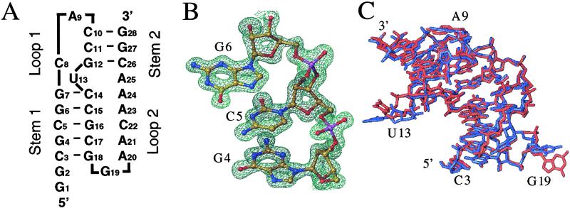
Many pathogenic viruses use programmed -1 ribosomal frameshifting to regulate translation of their structural and enzymatic proteins from polycistronic mRNAs. Frameshifting is commonly stimulated by a pseudoknot located downstream from a slippery sequence, the latter positioned at the ribosomal A and P sites. We report here the structures of two crystal forms of the frameshifting RNA pseudoknot from beet western yellow virus at resolutions of 1.25 and 2.85 A. Because of the very high resolution of 1.25 A, ten mono- and divalent metal ions per asymmetric unit could be identified, giving insight into potential roles of metal ions in stabilizing the pseudoknot. A magnesium ion located at the junction of the two pseudoknot stems appears to play a crucial role in stabilizing the structure. Because the two crystal forms exhibit mostly unrelated packing interactions and local crystallographic disorder in the high-resolution form was resolvable, the two structures offer the most detailed view yet of the conformational preference and flexibility of an RNA pseudoknot.


Figures

Figure 1
Figure 1
Sequence of the RNA construct and quality and conformational variation of the crystal structure as a function of the lattice. (A) Secondary structure diagram of the frameshifting pseudoknot from BWYV (residues C3–G28). (B) View of the final Fourier 2FoFc sum electron density in the trigonal crystal form at 1.25 Å resolution and contoured at 2σ (= 1 e/Å3) around residues G4, C5, and G6 from S1. RNA atoms are colored yellow, red, blue, and magenta for carbon, oxygen, nitrogen, and phosphorus, respectively. (C) Overall superposition of the pseudoknot structures in the trigonal (blue) and cubic (red) crystal forms.
Figure 2
Figure 2
Packing modes in the two crystal forms of the pseudoknot from BWYV. Views of selected lattice interactions in the (A) trigonal and (B) cubic crystal forms. Nucleotides belonging to neighboring molecules are colored differently. In both lattices, a 2-fold rotation axis positioned roughly perpendicular to the plane of projection relates symmetry mates. Alternative conformations of nucleotides G1 and U13 observed in the trigonal crystal form are indicated with thick and thin bonds and hydrogen bonds are thin dashed lines.
Figure 3
Figure 3
Coordination modes of metal ions in the trigonal crystal form. (A) View of individual magnesium (15) and monovalent ions (ref. ; four Na+ with full occupancy and two K+ with occupancy 0.5) colored gold and magenta, respectively, surrounding each pseudoknot molecule. All metal ions are engaged in either direct or water mediated contacts to the RNA. The backbone and base pair portions of stem regions are colored blue and yellow, respectively. L1 and L2 residues are colored red and green, respectively, and the base portions of U13 and A25 are highlighted in purple. Alternative conformations of G1, U13, and A23 and surrounding regions are indicated with thin cyan lines. (B) Stereo view of magnesium hexahydrates Mg45 and Mg52 in the major groove of S1. (C) Magnesium hexahydrate Mg38 in the major groove of S2. (D) Sodium ions Na61 and Na63 in the minor groove of S1. RNA atoms are colored yellow, red, blue, and magenta for carbon, oxygen, nitrogen, and phosphorus, respectively, a symmetry related molecule and magnesium ion are colored green (Fig. 3C), hydrogen bonds and coordination spheres of metal ions are indicated with thin dashed lines and selected residues are labeled.

References

    1. Farabaugh P J. Microbiol Rev. 1996;60:103–134. - PMC - PubMed
    1. Gesteland R F, Atkins J F. Annu Rev Biochem. 1996;65:741–768. - PubMed
    1. Giedroc D P, Theimer C A, Nixon P L. J Mol Biol. 2000;298:167–185. - PMC - PubMed
    1. Jacks T, Power M D, Masiarz F R, Lucius P A, Barr P J, Varmus H E. Nature (London) 1988;331:280–283. - PubMed
    1. Parkin N T, Chamorro M, Varmus H E. J Virol. 1992;66:5147–5151. - PMC - PubMed

Publication types