. 2002 Apr 2;99(7):4586-91.
doi: 10.1073/pnas.072647999. Epub 2002 Mar 19.

Impaired cardiac hypertrophic response in Calcineurin Abeta -deficient mice

Affiliations

Impaired cardiac hypertrophic response in Calcineurin Abeta -deficient mice

Orlando F Bueno et al. Proc Natl Acad Sci U S A. .

Abstract

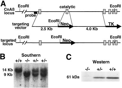
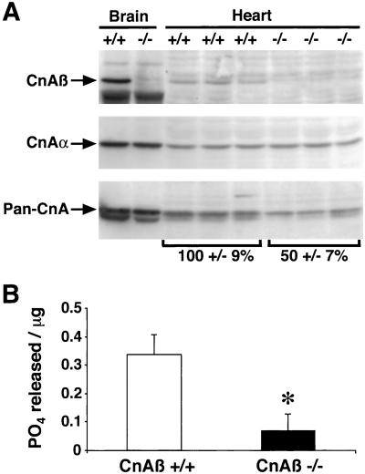
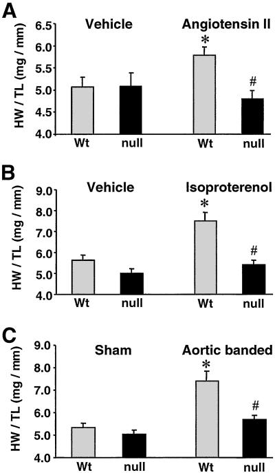
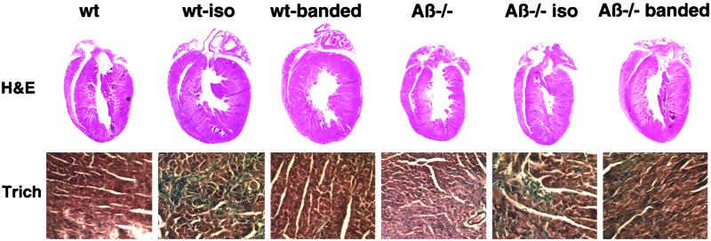
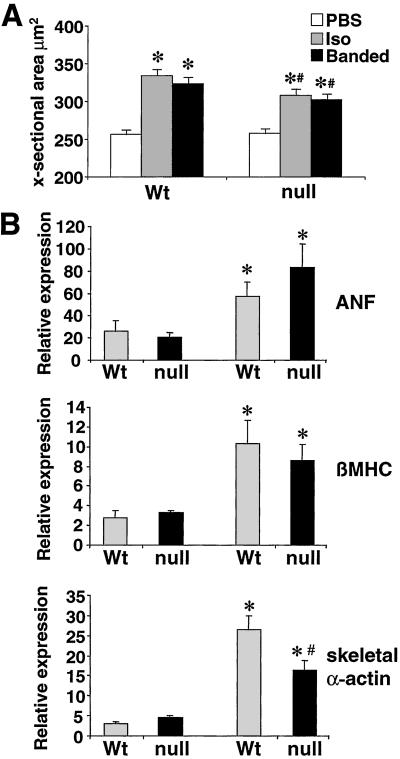
Calcineurin is a calcium-calmodulin-regulated, serine-threonine phosphatase that functions as a key inducer of stress responsive gene expression in multiple cell types through a direct activation of nuclear factor of activated T cells and myocyte enhancer factor 2 transcription factors. In cardiomyocytes, calcineurin signaling has been implicated in the regulation of the hypertrophic response caused by pressure overload or neuroendocrine stimulation. Three separate genes encode the catalytic subunit of calcineurin in mammalian cells, CnAalpha, CnAbeta, and CnAgamma. To evaluate the necessary function of calcineurin as a hypertrophic regulatory factor, the CnAbeta gene was disrupted in the mouse. CnAbeta-deficient mice were viable, fertile, and overtly normal well into adulthood, but displayed a 80% decrease in calcineurin enzymatic activity in the heart that was associated with a 12% reduction in basal heart size. CnAbeta-deficient mice were dramatically impaired in their ability to mount a productive hypertrophic response induced by pressure overload, angiotensin II infusion, or isoproterenol infusion. Analysis of marker genes associated with the hypertrophic response revealed a partial defect in the molecular program of hypertrophy. Collectively, these data solidify the hypothesis that calcineurin functions as a central regulator of the cardiac hypertrophic growth response in vivo.


Figures

Figure 1
Figure 1
Targeted disruption of the CnAβ gene. (A) Schematic representation of the CnAβ genomic locus, the targeting construct, and the result of homologous recombination that deletes the exon encoding the catalytic domain. (B) Southern blot analysis was performed using EcoRI-digested genomic DNA from four littermates obtained from CnAβ heterozygotes crosses, which show a wild-type (14 kb) and targeted fragment (9 Kb) of the predicted size. (C) Western blot analysis for CnAβ protein from brain extracts of wild-type, heterozygous, and null mice.
Figure 2
Figure 2
Analysis of cardiac calcineurin protein/activity from wild-type and CnAβ−/− mice. (A) Protein extracts from 6-week-old mice of either genotype were prepared for Western blotting and analyzed using antibodies that recognize either all catalytic subunit gene isoforms (Pan-CnA), or specific isoforms (CnAβ or CnAα). (B) Cardiac extracts from wild-type and CnAβ−/− mice were assayed for calcineurin activity (okadaic acid-resistant and EGTA-sensitive phosphatase activity). ∗, P < 0.05.
Figure 3
Figure 3
Quantitation of the cardiac hypertrophic response in CnAβ−/− mice. (A) Heart weight/tibia length ratios after 14 days of AngII infusion (432 μg/kg/day) in 8-week-old CnAβ+/+ and CnAβ−/− mice (n = 6 mice in each cohort). (B) Heart weight/tibia length ratios after 14 days of Iso infusion (60 mg/kg/day) in 8-week-old CnAβ+/+ and CnAβ−/− mice (n = 6 mice in each cohort). (C) Heart weight/tibia length ratios after 14 days of pressure overload induced by stenosis of the abdominal aorta in 8-week-old CnAβ+/+ and CnAβ−/− mice (n = 5 banded mice and 6 sham mice for both wild-type and CnAβ−/−). ∗, P < 0.05 versus wild-type vehicle or sham group; #, P < 0.05 versus wild-type treated or banded mice; Wt, wild type.
Figure 4
Figure 4
Cardiac histological analysis of wild-type and CnAβ−/− mice subjected to 14 days of Iso infusion or pressure overload. (Upper) Macroscopic hematoxylin/eosin-stained histological section of hearts demonstrate a noticeable increase in heart size in wild-type mice, but not CnAβ−/− mice. (Lower) Microscopic analysis of trichrome-stained histological sections demonstrate comparable myofibrillar organization and interstitial fibrosis in wild-type (wt) and CnAβ−/− mice treated with Iso or by pressure overload.
Figure 5
Figure 5
Myfibrillar cross-sectional areas and RNA dot blot analysis of hypertrophy associated genes from wild-type (Wt) and CnAβ−/− hearts. (A) Approximately 150 fibers were measured from each of the indicated groups to qunatify myofiber cross-sectional areas (∗, P < 0.05 versus unstimulated; #, P < 0.05 versus wild-type Iso-infused or banded). (B) Cardiac RNA levels quantified by dot blotting from mice subjected to 14 days of AngII infusion (432 μg/kg/day). RNA levels of atrial natriuretic factor (ANF), β-myosin heavy chain (βMHC), and skeletal α-actin, were analyzed (n = 4 each). Data are expressed as the percent of glyceraldehyde 3-phosphate dehydrogenase levels (∗, P < 0.05 versus vehicle-treated; #, P < 0.05 versus wild-type AngII-infused mice).

References

    1. Crabtree G R. Cell. 1999;96:611–614. - PubMed
    1. Klee C B, Ren H, Wang X. J Biol Chem. 1998;273:13367–13370. - PubMed
    1. Rusnak F, Mertz P. Physiol Rev. 2000;80:1483–1521. - PubMed
    1. Taigen T, De Windt L J, Lim H W, Molkentin J D. Proc Natl Acad Sci USA. 2000;97:1196–1201. - PMC - PubMed
    1. Haq S, Choukroun G, Lim H, Tymitz K M, del Monte F, Gwathmey J, Grazette L, Michael A, Hajjar R, Force T, Molkentin J D. Circulation. 2001;103:670–677. - PubMed

Publication types