. 2002 Mar 19;99(6):3557-62.
doi: 10.1073/pnas.062415899.

Mutation of the myosin converter domain alters cross-bridge elasticity

Affiliations

Mutation of the myosin converter domain alters cross-bridge elasticity

Jan Köhler et al. Proc Natl Acad Sci U S A. .

Abstract

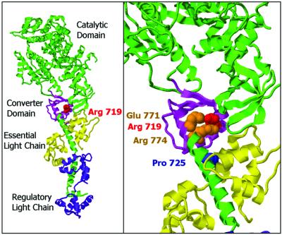
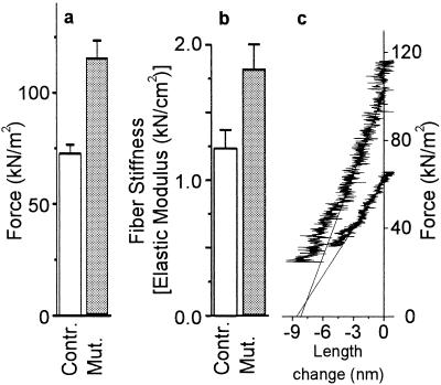
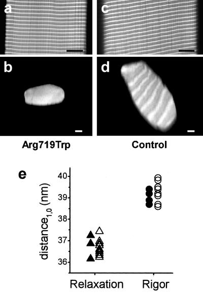
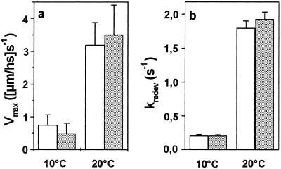
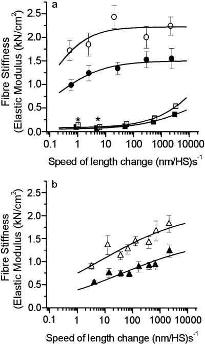
Elastic distortion of a structural element of the actomyosin complex is fundamental to the ability of myosin to generate motile forces. An elastic element allows strain to develop within the actomyosin complex (cross-bridge) before movement. Relief of this strain then drives filament sliding, or more generally, movement of a cargo. Even with the known crystal structure of the myosin head, however, the structural element of the actomyosin complex in which elastic distortion occurs remained unclear. To assign functional relevance to various structural elements of the myosin head, e.g., to identify the elastic element within the cross-bridge, we studied mechanical properties of muscle fibers from patients with familial hypertrophic cardiomyopathy with point mutations in the head domain of the beta-myosin heavy chain. We found that the Arg-719 --> Trp (Arg719Trp) mutation, which is located in the converter domain of the myosin head fragment, causes an increase in force generation and fiber stiffness under isometric conditions by 48-59%. Under rigor and relaxing conditions, fiber stiffness was 45-47% higher than in control fibers. Yet, kinetics of active cross-bridge cycling were unchanged. These findings, especially the increase in fiber stiffness under rigor conditions, indicate that cross-bridges with the Arg719Trp mutation are more resistant to elastic distortion. The data presented here strongly suggest that the converter domain that forms the junction between the catalytic and the light-chain-binding domain of the myosin head is not only essential for elastic distortion of the cross-bridge, but that the main elastic distortion may even occur within the converter domain itself.


Figures

Figure 1
Figure 1
Ribbon diagram of the structure of the scallop striated muscle subfragment-1 (17) showing the position of the FHC mutation studied here. (Left) Heavy chain of S1 (catalytic domain pointing to the upper left) and the long α-helix representing the lever arm are in green, light chains in magenta and yellow, the converter domain in light violet. The converter domain encompasses residues Phe-707 to Arg-774 (17). Arg-719 is displayed space-filling in red. The crystal structure shown here indicates the very close proximity of the mutated residue and the first turns of the α-helix forming the core of the light-chain-binding domain. (Right) Close-up view of the converter area highlighting two residues of the long α-helix (Glu-771 and Arg-774, space-filling in orange) that are in close contact with Arg-719, which has been described to be conserved in all heavy-chain isoforms (17). Pro-725 of the converter is shown space-filling in blue to illustrate the part of the converter that is close to the essential light chain. (Figure prepared with RasMol.)
Figure 2
Figure 2
Effect of the Arg719Trp mutation on mechanical parameters of single fibers from soleus muscle. (a) Active isometric force [19 control fibers (open bars) and 18 fibers with mutated myosin (gray bars)] at 10°C. (b) Isometric fiber stiffness [14 control fibers (open bars) and 9 fibers with mutated myosin (gray bars)] at 10°C. (c) Original records of stiffness measurements during releases starting from isometric steady state. Force is plotted vs. change in sarcomere length recorded during the imposed release; speed of releases ≈2.2 × 10³ (nm/half-sarcomere) s−1. Upper plot, fiber with Arg719Trp mutation; lower plot, control fiber; solid lines, linear least squares fits to the linear part of the plots (some 3 nm/half-sarcomere for the control fiber and 4 nm/half-sarcomere for the fiber with Arg719Trp mutation). Here, the rather similar intercept of these plots with the x axis (y0 value) shows that approximately the same-length change (release) is necessary to drop active force to zero. Note that, on average, the length change necessary to drop active force to zero was about 10% higher for fibers with mutated myosin than for controls.
Figure 3
Figure 3
Confocal images and x-ray diffraction data to ensure precise measurement of fiber cross-sections and normal content of myofibrils and myofilaments in both fibers with mutant myosin and control fibers. (a and c) Longitudinal optical sections through the core of the fibers; (b and d) optical cross-sections of the same fibers. (Left) Fiber with mutated myosin; (Right) control fiber. (Scale bars: 10 μm.) Fibers were labeled with rhodamine-phalloidin, which under the conditions used here binds to the actin filaments in the overlap region and near the Z-line. The light and dark pattern of the cross-sections arises form cutting through unlabeled and labeled regions of the sarcomere. (e) Distance of the two innermost equatorial reflections (d1,0) in x-ray diffraction patterns obtained in rigor and relaxation from fibers with mutated myosin (open symbols) and control fibers (filled symbols).
Figure 4
Figure 4
Mechanical parameters sensitive to cross-bridge turnover kinetics. Gray bars, muscle fibers with mutated myosin; open bars, control fibers. (a) Maximum unloaded shortening velocity (vmax) of six fibers each, and (b) rate constant of force redevelopment (kredev) of 21 control fibers and 20 fibers with mutated myosin. Neither vmax nor kredev are affected by the mutation.
Figure 5
Figure 5
Stiffness of muscle fibers with mutated myosin and of control fibers. (a) Stiffness in rigor (filled circles, 7 control fibers; open circles, 6 fibers with mutation) and under relaxing conditions (filled squares, 14 control fibers; open squares, 10 fibers with mutation), both measured at 5°C. (b) Active isometric stiffness (filled triangles, 14 control fibers; open triangles, 9 fibers with mutation) at 10°C. Lines represent parts of sigmoidal fits to the data points. Stiffness in rigor and relaxation was measured at 5°C to enhance relaxed stiffness. Previous measurements showed an approximately 10% and 15% decrease of relaxed stiffness when raising the temperature from 5°C to 10°C or to 20°C, respectively, whereas rigor stiffness was not affected by these temperature changes. *Difference not statistically significant (P > 0.05).

Similar articles

Cited by

References

    1. Rayment I, Holden H M, Whittaker M, Yohn C B, Lorenz M, Holmes K C, Milligan R A. Science. 1993;261:58–65. - PubMed
    1. Lowey S, Waller G S, Trybus K M. Nature (London) 1993;365:454–456. - PubMed
    1. Whittaker M, Wilson-Kubalek E M, Smith J E, Faust L, Milligan R A, Sweeney H L. Nature (London) 1995;378:748–751. - PubMed
    1. Uyeda T Q, Abramson P D, Spudich J A. Proc Natl Acad Sci USA. 1996;93:4459–4464. - PMC - PubMed
    1. Dominguez R, Freyzon Y, Trybus K M, Cohen C. Cell. 1998;94:559–571. - PubMed

Publication types

MeSH terms

LinkOut - more resources