. 2002 Mar 19;99(6):3794-9.
doi: 10.1073/pnas.062055999.

Negative regulation of Lck by Cbl ubiquitin ligase

Affiliations

Negative regulation of Lck by Cbl ubiquitin ligase

Navin Rao et al. Proc Natl Acad Sci U S A. .

Abstract

The Cbl-family ubiquitin ligases function as negative regulators of activated receptor tyrosine kinases by facilitating their ubiquitination and subsequent targeting to lysosomes. Cbl associates with the lymphoid-restricted nonreceptor tyrosine kinase Lck, but the functional relevance of this interaction remains unknown. Here, we demonstrate that T cell receptor and CD4 coligation on human T cells results in enhanced association between Cbl and Lck, together with Lck ubiquitination and degradation. A Cbl(-/-) T cell line showed a marked deficiency in Lck ubiquitination and increased levels of kinase-active Lck. Coexpression in 293T cells demonstrated that Lck kinase activity and Cbl ubiquitin ligase activity were essential for Lck ubiquitination and negative regulation of Lck-dependent serum response element-luciferase reporter activity. The Lck SH3 domain was pivotal for Cbl-Lck association and Cbl-mediated Lck degradation, with a smaller role for interactions mediated by the Cbl tyrosine kinase-binding domain. Finally, analysis of a ZAP-70-deficient T cell line revealed that Cbl inhibited Lck-dependent mitogen-activated protein kinase activation, and an intact Cbl RING finger domain was required for this functional effect. Our results demonstrate a direct, ubiquitination-dependent, negative regulatory role of Cbl for Lck in T cells, independent of Cbl-mediated regulation of ZAP-70.


Figures
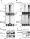
Figure 1
Figure 1
Lck associates with Cbl and is ubiquitinated upon TCR stimulation in SPF1 T cells. (A) Resting CD4+ human SPF1 T cells that had been incubated for 12 h in the absence of IL-2 were stimulated by cross-linking with no antibody (−), control antibody, or anti-CD3/4 antibodies for 10 min at 37°C and then lysed in SDS lysis buffer. Anti-Lck IPs from 1-mg aliquots of lysates was serially probed with an anti-Ub (Top) and anti-Lck antibody (Bottom). (B) SPF1 T cells were treated with 50 μM MG132 (+) or dimethyl sulfoxide vehicle control (−) for 3 h before stimulation for 10 min. IPs were performed and immunoblotted as above. (C) Equal amounts of protein lysates from B were serially probed with anti-Lck and anti-MAPK antibodies. (D) SPF1 T cells were stimulated for 5 min., lysed in Triton lysis buffer, and anti-Lck and isotype matched control IPs were immunoblotted with anti-Cbl (Top) or anti-Lck antibody (Bottom).
Figure 2
Figure 2
Cbl−/− T cells have increased levels of kinase-active Lck. (A) Cbl−/− and Cbl+/+ T cells were lysed in RIPA buffer and equal amounts (50 μg) of protein lysates were immunoblotted with anti-Cbl (top panel), anti-Lck (second panel) and anti-MAPK (bottom panel) antibodies. Anti-Lck IPs from 250 μg of protein lysate was immunoblotted with anti-phospho-Lck (third panel). (B) Anti-Lck or isotype matched control IPs from lysates used in A, or a positive control lysate from transiently transfected 293T cells, were subjected to in vitro kinase assays and the incorporation of 32P signal of [γ32P]ATP into a synthetic Raytide substrate (+) or negative control substrate (−) was quantified. Results are expressed as the mean ± 1 SD of three replicates.
Figure 3
Figure 3
Impaired Lck ubiquitination in Cbl−/− T cells. Cbl−/− and Cbl+/+ T cells were incubated with 10 μM Lactacystin (+) or dimethyl sulfoxide control (−) as well as 0.1 mM orthovanadate for 5 h and then lysed in RIPA buffer. Anti-Lck IPs from 1 mg aliquots of lysate were immunoblotted with anti-Ub antibody (Top), followed by anti-Lck antibody (Middle). Equal aliquots (30 μg) of cell lysates were immunoblotted with anti-Lck antibody (Bottom).
Figure 4
Figure 4
Cbl-dependent ubiquitination of Lck in 293T cells in dependent on the Cbl RING finger domain and Lck kinase activity. (A) 293T cells were transfected with plasmids encoding HA-Ub (5 μg), Lck (0.2 μg), and 3 μg of GFP-Cbl (WT), GFP-Cbl-C3AHN RING finger mutant, or a GFP control (−). Cells were lysed in RIPA buffer, and anti-Lck IPs from 800-μg aliquots of lysate protein were immunoblotted with anti-HA antibody (Top). Equal aliquots (30 μg) of cell lysates were immunoblotted with anti-Lck antibody (Middle) followed by anti-GFP antibody (Bottom). Control GFP is not included in the blot. (B) 293T cells were transfected with plasmids encoding HA-Ub (5 μg), Lck (WT), kinase active (Y505F), and kinase dead (R273A) (0.2 μg each), and GFP-Cbl or a GFP control (−) (3 μg). Cells were lysed in RIPA buffer, and immunoblots of anti-Lck IPs were performed as in A.
Figure 5
Figure 5
Relative contribution of Lck SH2 and SH3 domains and the Cbl TKB domain toward Cbl-mediated Lck degradation. 293T cells were transfected with plasmids encoding HA-Cbl (1 μg) and Lck (WT), SH2 (R154K), SH3 (W97A), or double mutants (R154K/W97A) (0.5 μg each). Cells were lysed in Triton lysis buffer and anti-Lck IPs from 1-mg aliquots of lysate protein were immunoblotted with anti-HA antibody (Top) and anti-Lck antibody (Upper Middle). Aliquots (20 μg) of lysate protein were immunoblotted with anti-HA antibody (Lower Middle). For degradation, 293T cells were transfected as above, but with 0.2 μg of the indicated Lck plasmids and lysed with RIPA buffer. 20 μg aliquots of lysate protein were immunoblotted with anti-Lck antibody (Bottom). Lck protein levels were quantified by densitometry and are expressed relative to each Lck protein in the absence of coexpressed Cbl.
Figure 6
Figure 6
The RING finger domain is required for Cbl-dependent negative regulation of Lck. (A) 293T cells were transfected with plasmids encoding the SRE-luciferase reporter (5 μg) and the indicated combinations of Lck (0.15 μg), HA-Cbl, HA-Cbl-C3AHN and HA-Cbl-70Z (1 μg) or pAlterMAX vector (−). Luciferase activity was expressed relative to activity of lysates transfected with the reporter in the absence of Lck or Cbl. Results represent the mean ± one SD of five replicate transfections. (B) Jurkat-derived ZAP-70-deficient p116-T cells, were transfected with 15 μg of plasmid DNA encoding HA-Cbl, HA-Cbl-C3AHN, or pAlterMAX vector (−). Cells were either left unstimulated or stimulated for the indicated times with anti-CD3 antibody before lysis in RIPA buffer. Equal aliquots of cell lysates (25 μg) were subjected to anti-phospho-MAPK (Top), anti-MAPK (Upper Middle), anti-Lck (Lower Middle), and anti-HA (Bottom) immunoblotting.

Similar articles

Cited by

References

    1. Latour S, Veillette A. Curr Opin Immunol. 2001;13:299–306. - PubMed
    1. Cheng A M, Negishi I, Anderson S J, Chan A C, Bolen J, Loh D Y, Pawson T. Proc Natl Acad Sci USA. 1997;94:9797–9801. - PMC - PubMed
    1. Gupta S, Weiss A, Kumar G, Wang S, Nel A. J Biol Chem. 1994;269:17349–17357. - PubMed
    1. Molina T J, Kishihara K, Siderovski D P, van Ewijk W, Narendran A, Timms E, Wakeham A, Paige C J, Hartmann K U, Veillette A, et al. Nature (London) 1992;357:161–164. - PubMed
    1. Groves T, Smiley P, Cooke M P, Forbush K, Perlmutter R M, Guidos C J. Immunity. 1996;5:417–428. - PubMed

Publication types

MeSH terms

LinkOut - more resources