. 2002 Mar 19;99(6):3812-7.
doi: 10.1073/pnas.052707499.

Prions in skeletal muscle

Affiliations

Prions in skeletal muscle

Patrick J Bosque et al. Proc Natl Acad Sci U S A. .

Abstract

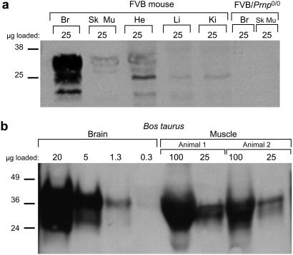
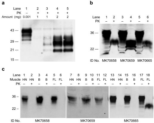
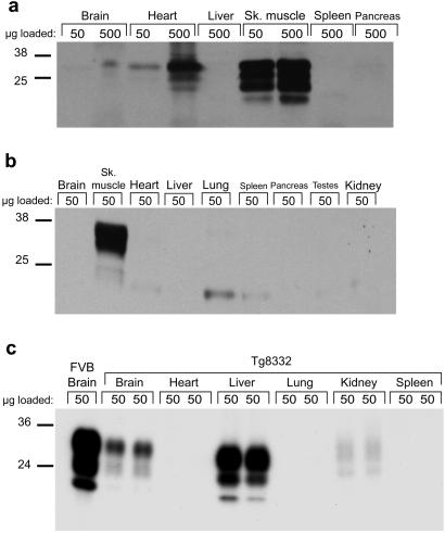
Considerable evidence argues that consumption of beef products from cattle infected with bovine spongiform encephalopathy (BSE) prions causes new variant Creutzfeldt-Jakob disease. In an effort to prevent new variant Creutzfeldt-Jakob disease, certain "specified offals," including neural and lymphatic tissues, thought to contain high titers of prions have been excluded from foods destined for human consumption [Phillips, N. A., Bridgeman, J. & Ferguson-Smith, M. (2000) in The BSE Inquiry (Stationery Office, London), Vol. 6, pp. 413-451]. Here we report that mouse skeletal muscle can propagate prions and accumulate substantial titers of these pathogens. We found both high prion titers and the disease-causing isoform of the prion protein (PrP(Sc)) in the skeletal muscle of wild-type mice inoculated with either the Me7 or Rocky Mountain Laboratory strain of murine prions. Particular muscles accumulated distinct levels of PrP(Sc), with the highest levels observed in muscle from the hind limb. To determine whether prions are produced or merely accumulate intramuscularly, we established transgenic mice expressing either mouse or Syrian hamster PrP exclusively in muscle. Inoculating these mice intramuscularly with prions resulted in the formation of high titers of nascent prions in muscle. In contrast, inoculating mice in which PrP expression was targeted to hepatocytes resulted in low prion titers. Our data demonstrate that factors in addition to the amount of PrP expressed determine the tropism of prions for certain tissues. That some muscles are intrinsically capable of accumulating substantial titers of prions is of particular concern. Because significant dietary exposure to prions might occur through the consumption of meat, even if it is largely free of neural and lymphatic tissue, a comprehensive effort to map the distribution of prions in the muscle of infected livestock is needed. Furthermore, muscle may provide a readily biopsied tissue from which to diagnose prion disease in asymptomatic animals and even humans.


Figures

Figure 1
Figure 1
PrPC expression in mammalian muscle. (a) Western blot depicting the level of PrPC expression in various tissues of an FVB mouse. The highest level of PrPC is in brain (Br), but distinct bands corresponding to fully glycosylated PrPC are also seen in lanes with skeletal muscle (Sk Mu) and heart (He) homogenates. Faint bands apparently representing partially glycosylated PrPC are found in lanes bearing liver (Li) and kidney (Ki) homogenates. No bands are seen in tissues from an FVB/Prnp0/0 mouse. The blot was probed with the R073 polyclonal antiserum. (b) Western blots comparing PrPC expression level in the brain and muscle of Bos taurus. Serial dilutions of brain homogenate from a single animal were compared with dilutions of muscle homogenates from two animals obtained from a different source. The amount of PrPC found in 25 μg of muscle homogenate is slightly greater than that seen in 1.3 μg of brain homogenate; therefore, we conclude that PrPC expression in muscle is ≈5–10% of that in brain. The PrPC derived from muscle in this blot migrated faster than that derived from brain because the muscle is from cattle bearing a 5-octarepeat PRNP allele, whereas brain is derived from an animal with the 6-octarepeat allele (50).
Figure 2
Figure 2
Protease-resistant PrPSc in muscle of RML- and Me7-infected CD-1 and FVB mice. (a) Lane 1 contains untreated brain homogenate from an uninoculated CD-1 mouse. All other lanes contain insoluble fractions from PK-digested homogenates of CD-1 mouse muscle. No protease-resistant PrPSc was found in the muscle of uninoculated mice (lane 2), whereas PrPSc was readily detectable in the muscle of mice showing signs of neurologic disease 132 days after intracerebral inoculation with RML prions (lanes 4 and 5). For comparison, brain homogenate from a mouse showing signs of disease was diluted 1:1000 into a muscle homogenate from an uninoculated mouse (lane 3). Amounts loaded are expressed as equivalents of total protein (in mg) of homogenate, before proteinase digestion and precipitation. Note that lanes 4 and 5 were loaded with the product of twice as much original homogenate as lanes 2 and 3. (b) Hindlimb skeletal muscles of three ill FVB mice inoculated with Me7 prions were homogenized and subjected to analysis. These mice showed typical clinical signs of disease at 138 days (MK70665) and 146 days (MK70658, MK70659) after intracerebral inoculation with Me7 prions. Samples were digested with PK (lanes 2, 4, and 6) or undigested (lanes 1, 3, and 5). Two milligrams of PK-digested muscle homogenates and about 60 μg of undigested muscle homogenates were used for analysis. (c) Muscle tissue from the head-neck (HN), back (B), and forelimb (FL) of the mice analyzed in b were collected at the time that the hindlimb muscle was collected. Lanes were loaded with PK-digested (even-numbered lanes) and undigested muscle homogenates (odd-numbered lanes). The analytical method was the same as that described in b. All blots were probed with the HuM-D13 Ab.
Figure 3
Figure 3
Western blots depicting the distribution of PrPC expression in Tg mice. (a) Tg(α-actin-MoPrP)6906/Prnp0/0 mouse probed with the polyclonal Ab R073. PrPC expression in muscle is about 8-fold higher than in wt mouse brain (not shown). (b) Tg(MCK-SHaPrP)8775/Prnp0/0 mouse probed with 3F4, a mAb that recognizes hamster PrPC. PrPC expression is detectable only in skeletal muscle (Sk. muscle). A longer exposure of this blot (not shown) revealed trace expression in cardiac muscle, but no PrPC was detected in any other organ. The low molecular weight bands, seen most prominently in lane 5 (Lung), were also seen when the anti-mouse Ig secondary Ab was used without prior incubation with 3F4. (c) Homogenates of organs from two Tg(TTR-MoPrP) 8332/Prnp0/0 mice compared with FVB brain. The blot was probed with R073. PrPC expression in liver is lower than in FVB brain but higher than in the brain of mice of this Tg line. A very small amount of PrPC is expressed in the kidney. Glycosylated forms of PrPC from the liver migrate slightly faster than the corresponding glycoforms from the brain of these same mice or of FVB mice.

Similar articles

Cited by

References

    1. Prusiner S B. Science. 1982;216:136–144. - PubMed
    1. Büeler H, Aguzzi A, Sailer A, Greiner R-A, Autenried P, Aguet M, Weissmann C. Cell. 1993;73:1339–1347. - PubMed
    1. Prusiner S B, Groth D, Serban A, Koehler R, Foster D, Torchia M, Burton D, Yang S-L, DeArmond S J. Proc Natl Acad Sci USA. 1993;90:10608–10612. - PMC - PubMed
    1. Eklund C M, Kennedy R C, Hadlow W J. J Infect Dis. 1967;117:15–22. - PubMed
    1. Hadlow W J, Kennedy R C, Race R E, Eklund C M. Vet Pathol. 1980;17:187–199. - PubMed

Publication types

MeSH terms