. 2002 Mar 19;99(6):3818-23.
doi: 10.1073/pnas.062605899.

Profound defects in pancreatic beta-cell function in mice with combined heterozygous mutations in Pdx-1, Hnf-1alpha, and Hnf-3beta

Affiliations

Profound defects in pancreatic beta-cell function in mice with combined heterozygous mutations in Pdx-1, Hnf-1alpha, and Hnf-3beta

David Q Shih et al. Proc Natl Acad Sci U S A. .

Abstract

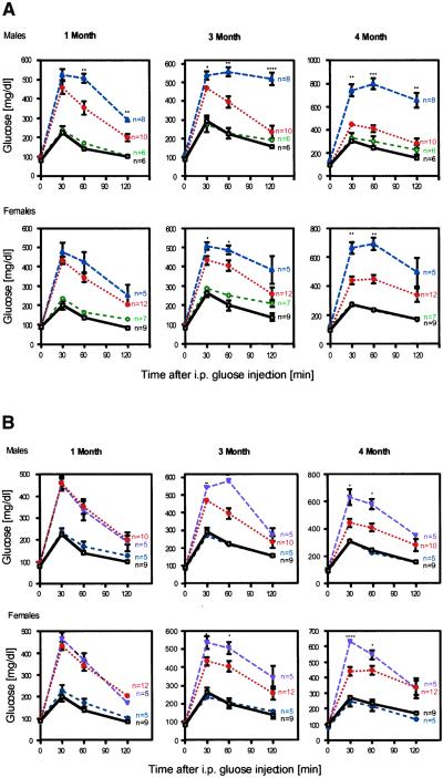
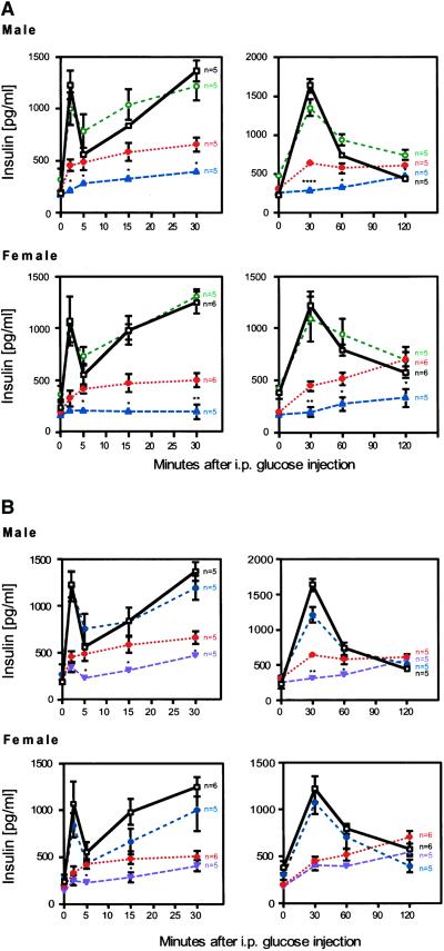
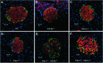
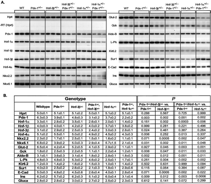
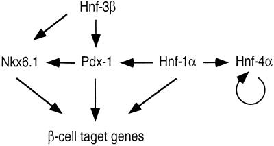
Defects in pancreatic beta-cell function contribute to the development of type 2 diabetes, a polygenic disease that is characterized by insulin resistance and compromised insulin secretion. Hepatocyte nuclear factors (HNFs) -1alpha, -3beta, -4alpha, and Pdx-1 contribute in the complex transcriptional circuits within the pancreas that are involved in beta-cell development and function. In mice, a heterozygous mutation in Pdx-1 alone, but not Hnf-1alpha(+/-), Hnf-3beta(+/-), or Hnf-4alpha(+/-), causes impaired glucose-stimulated insulin secretion in mice. To investigate the possible functional relationships between these transcription factors on beta-cell activity in vivo, we generated mice with the following combined heterozygous mutations: Pdx-1(+/-)/Hnf-1alpha(+/-), Pdx-1(+/-)/Hnf-3beta(+/-), Pdx-1(+/-)/Hnf-4alpha(+/-), Hnf-1alpha(+/-)/Hnf-4alpha(+/-), and Hnf-3beta(+/-)/Hnf-4alpha(+/-). The greatest loss in function was in combined heterozygous null alleles of Pdx-1 and Hnf-1alpha (Pdx-1(+/-)/Hnf-1alpha(+/-)), or Pdx-1 and Hnf-3beta (Pdx-1(+/-)/Hnf-3beta(+/-)). Both double mutants develop progressively impaired glucose tolerance and acquire a compromised first- and second-phase insulin secretion profile in response to glucose compared with Pdx-1(+/-) mice alone. The loss in beta-cell function in Pdx-1(+/-)/Hnf-3beta(+/-) mice was associated with decreased expression of Nkx-6.1, glucokinase (Gck), aldolase B (aldo-B), and insulin, whereas Nkx2.2, Nkx-6.1, Glut-2, Gck, aldo-B, the liver isoform of pyruvate kinase, and insulin expression was reduced in Pdx-1(+/-)/Hnf-1alpha(+/-) mice. The islet cell architecture was also abnormal in Pdx-1(+/-)/Hnf-3beta(+/-) and Pdx-1(+/-)/Hnf-1alpha(+/-) mice, with glucagon-expressing cells scattered throughout the islet, a defect that may be connected to decreased E-cadherin expression. Our data suggest that functional interactions between key islet regulatory factors play an important role in maintaining islet architecture and beta-cell function. These studies also established polygenic mouse models for investigating the mechanisms contributing to beta-cell dysfunction in diabetes.


Figures

Figure 1
Figure 1
Progressive impairment of glucose homeostasis in Pdx-1+/−/Hnf-3β+/− and Pdx-1+/−/Hnf-1α+/− mice. IPGTT was performed in Wt (black, □), Hnf-1α+/− (green, ○), Pdx-1+/− (red, ⋄), Pdx-1+/−/Hnf-1α+/− (blue, ▵), and Pdx-1+/−/Hnf-3β+/− (purple,▿ ) littermates at 1, 3, and 4 months of age as described in Materials and Methods. (A) Significantly increased serum glucose concentrations were observed in male and female Pdx-1+/−/Hnf-1α+/− mice compared with Pdx-1+/− at 1, 3, and 4 months, respectively. (B) The IPGTTs of Pdx-1+/−/Hnf-3β+/− mice were indistinguishable from those of Pdx-1+/− littermates at 1 month but blood glucose levels increased in male and female mice after 3 months. A worsening of the phenotype was seen in all double heterozygous animals. *, P ≤ 0.05; **, P ≤ 0.01; ***, P ≤ 0.005; ****, P ≤ 0.0001.
Figure 2
Figure 2
Loss of first-phase and blunted second-phase insulin secretion in response to glucose. Glucose-stimulated insulin release was measured as described in Materials and Methods. (Left) Acute insulin secretion responses. (Right) Insulin levels over a 2-h period after glucose challenge. The data of Left and Right were collected independently and mice were allowed to recover for 1 week. Wt mice are shown in black (□), Hnf-1α+/− in green (○), Pdx-1+/− in red (⋄), Pdx-1+/−/Hnf-1α+/− in blue (▵), and Pdx-1+/−/Hnf-3β+/− in purple (▿). (A) Male and female Pdx-1+/−/Hnf-1α+/− mutant mice show a complete loss of acute-phase insulin response and a blunted second-phase insulin response. (B) Selective loss of acute-phase insulin secretion in Pdx-1+/−/Hnf-3β+/− mutant mice compared with Pdx-1+/− littermates. *, P ≤ 0.05; **, P ≤ 0.01; ***, P ≤ 0.005; ****, P ≤ 0.0001.
Figure 3
Figure 3
Pdx-1+/−/Hnf-3β+/− and Pdx-1+/−/Hnf-1α+/− mice exhibit altered islet morphology. Immunofluorescent confocal microscopy of pancreatic islets of Wt (A), Hnf-3β+/− (B), Hnf-1α+/− (C), Pdx-1+/− (D), Pdx-1+/−/Hnf-3β+/− (E), and Pdx-1+/−/Hnf-1α+/− (F) mice. Insulin-expressing cells are shown in red, glucagon-containing cells in green. Nuclei of respective cells are shown in blue. Islets from Pdx-1+/−/Hnf-3β+/− (E) and Pdx-1+/−/Hnf-1α+/− (F) mice show scattered α-cells throughout the islets. Pancreatic islet size was not significantly different in the different mutant mice.
Figure 4
Figure 4
(A) Steady-state mRNA levels of pancreatic islet enriched genes of wild-type (WT), Pdx-1+/−, Hnf-3β+/−, Hnf-1α+/−, Pdx-1+/−/Hnf-3β+/−, and Pdx-1+/−/Hnf-1α+/− mutant mice. The housekeeping gene hypoxanthine phosphoribosyltransferase (Hprt) was amplified to show that each sample contained similar amounts of mRNA. A lack of any amplification signal in reactions without reverse transcriptase (−RT) shows that genomic DNA did not contaminate the samples. Transcript levels were measured in three animals for each genotype by RT-PCR using [α-32P]dCTP. PCR products were separated by PAGE, and bands were visualized by autoradiography. (B) Quantitative measurements of gene expression were obtained by densitometry, and the mean of measurements are shown ± SD. The level of significance of the different comparisons (single- vs. double-heterozygous mutant) is shown on the right.
Figure 5
Figure 5
Regulation of pancreatic islet transcription factors. Strong synergistic effects exist in vivo between Pdx-1, Hnf-1α, and Hnf-3β. Hnf-1α and Hnf-3β both regulate the expression of Pdx-1 in vivo and in vitro. Hnf-4α expression is normal in Hnf-4α+/− mutant mice, suggesting an autoregulatory feedback loop. Furthermore, Hnf-4α does not regulate the expression of Hnf-1α or Pdx-1 in pancreatic islets. Gene expression analysis also indicates that Nkx6.1 gene transcript levels are regulated by Pdx-1.

Similar articles

Cited by

References

    1. Edlund H. Diabetes. 2001;50, Suppl. 1:S5–S9. - PubMed
    1. Shih, D. Q. & Stoffel, M. (2002) Curr. Diabetes Rep. 2, in press. - PubMed
    1. St-Onge L, Wehr R, Gruss P. Curr Opin Genet Dev. 1999;9:295–300. - PubMed
    1. Sander M, Sussel L, Conners J, Scheel D, Kalamaras J, Dela Cruz F, Schwitzgebel V, Hayes-Jordan A, German M. Development (Cambridge, UK) 2000;127:5533–5540. - PubMed
    1. Shim E Y, Woodcock C, Zaret K S. Genes Dev. 1998;12:5–10. - PMC - PubMed

Publication types

MeSH terms

LinkOut - more resources