. 2002 Mar 19;99(6):3938-43.
doi: 10.1073/pnas.062425699.

Chronic intracellular infection of alfalfa nodules by Sinorhizobium meliloti requires correct lipopolysaccharide core

Affiliations

Chronic intracellular infection of alfalfa nodules by Sinorhizobium meliloti requires correct lipopolysaccharide core

Gordon R O Campbell et al. Proc Natl Acad Sci U S A. .

Abstract

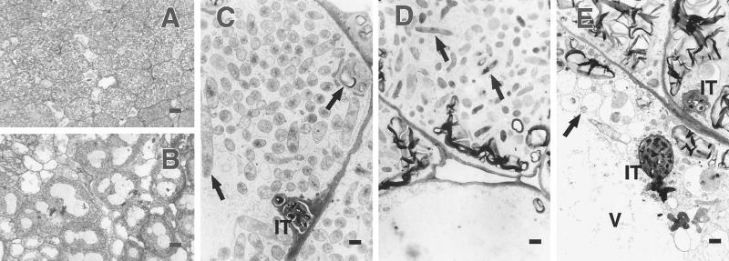
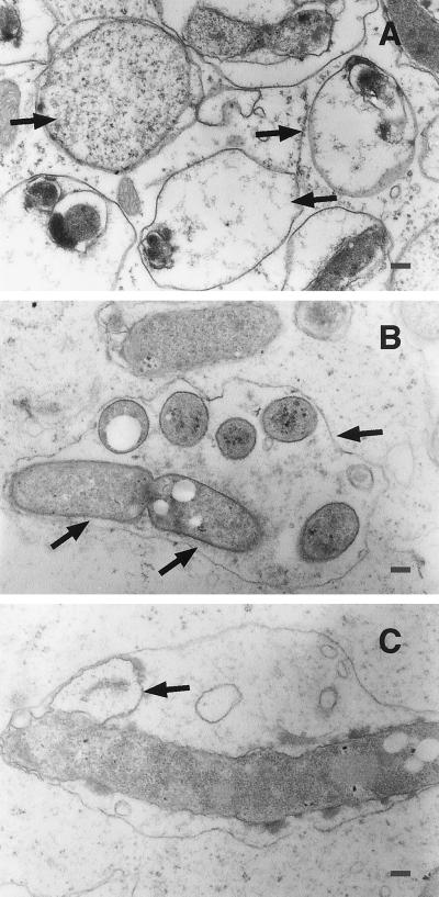
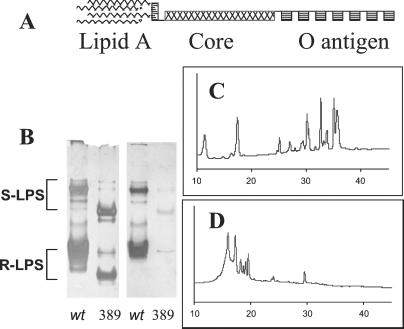
Our analyses of lipopolysaccharide mutants of Sinorhizobium meliloti offer insights into how this bacterium establishes the chronic intracellular infection of plant cells that is necessary for its nitrogen-fixing symbiosis with alfalfa. Derivatives of S. meliloti strain Rm1021 carrying an lpsB mutation are capable of colonizing curled root hairs and forming infection threads in alfalfa in a manner similar to a wild-type strain. However, developmental abnormalities occur in the bacterium and the plant at the stage when the bacteria invade the plant nodule cells. Loss-of-function lpsB mutations, which eliminate a protein of the glycosyltransferase I family, cause striking changes in the carbohydrate core of the lipopolysaccharide, including the absence of uronic acids and a 40-fold relative increase in xylose. We also found that lpsB mutants were sensitive to the cationic peptides melittin, polymyxin B, and poly-l-lysine, in a manner that paralleled that of Brucella abortus lipopolysaccharide mutants. Sensitivity to components of the plant's innate immune system may be part of the reason that this mutant is unable to properly sustain a chronic infection within the cells of its host-plant alfalfa.


Figures

Figure 1
Figure 1
Light micrographs (A and B) and electron micrographs (C–E) of region corresponding to nitrogen-fixing zone in nodules induced by the Rm1021 wild-type bacteria and by an lpsB mutant. (A) Cross section through the nitrogen-fixing region [Zone III as described by Vasse et al. (4)] of an alfalfa nodule infected with the wild-type S. meliloti strain Rm1021. (Bar = 18 μm.) (B) Cross section through region that would correspond to Zone III of an alfalfa nodule infected with an lpsB mutant. Cells contain many abnormally large vacuoles. (Bar = 18 μm.) (C) Plant cells packed with wild-type bacteroids (for example, left arrow) from a nodule infected by the wild-type S. meliloti strain Rm1021. An infection thread (IT) and an amyloplast (right arrow) are also shown. (Bar = 1.25 μm.) (D) Plant cells infected with an lpsB mutant. Bacteria are in various stages of degradation (arrows). (Bar = 1.25 μm.) (E) Plant cells from a nodule containing lpsB mutant bacteria. The bacteria are in a more degraded state, frequently being within enlarged membrane compartments (arrow). Infection threads are also present (IT). A large vacuole fills most of the cytoplasm of lower plant cell (V). (Bar = 1.25 μm.)
Figure 2
Figure 2
Intracellular lpsB mutant bacteria at a higher magnification in nodule cells from the region that would correspond to the nitrogen-fixing zone of a wild-type nodule. (A) Degrading bacteria within enlarged membrane compartments (right arrows). One bacterium has homogeneous staining in the cytoplasm similar to a bacteroid from the senescent zone (left arrow). (Bar = 0.28 μm.) (B) An enlarged membrane compartment (right arrow) containing several partially developed bacteroids. The elongated bacterium (bottom two arrows) appears to contain polyhydroxybutyrate granules (white circles). (Bar = 0.21 μm.) (C) An elongated bacterium, inside an enlarged membrane compartment, with a blebbing of bacterial outer membrane (arrow). (Bar = 0.17 μm.)
Figure 3
Figure 3
(A) Schematic of the LPS molecule showing the lipid A, the LPS core, and the O antigen. (B) Deoxycholic acid PAGE gel and Western blot of hot phenol-water purified LPS from wild-type and mutant LPS. R- and S-LPS are labeled accordingly. Both the smooth and rough LPS have run further in the mutant. LPS was blotted with polyclonal antibodies recognizing mostly the core region of LPS. The low intensity of the bands in the SmGC1 blot suggests that the core has been altered. (C) HPAEC-pulsed amperometric detection analysis of core oligosaccharides from R-LPS of wild-type S. meliloti and (D) the SmGC1 mutant. The mutant lacked all but one of the oligosaccharide peaks present in R-LPS core from the wild-type strain.

Similar articles

Cited by

References

    1. van Rhijn P, Vanderleyden J. Microbiol Rev. 1995;59:124–142. - PMC - PubMed
    1. Brewin N J. Annu Rev Cell Biol. 1991;7:191–226. - PubMed
    1. Hirsch A M, Long S R, Bang M, Haskins N, Ausubel F M. J Bacteriol. 1982;151:411–419. - PMC - PubMed
    1. Vasse J, deBilly F, Camut S, Truchet G. J Bacteriol. 1990;172:4295–4306. - PMC - PubMed
    1. Noel K D, Duelli D M. In: Prokaryotic Nitrogen Fixation: A Model System for Analysis of a Biological Process. Triplett E W, editor. Wymondham, U.K.: Horizon; 2000. pp. 415–431.

Publication types

MeSH terms

LinkOut - more resources