. 2002 Mar 19;99(6):3974-9.
doi: 10.1073/pnas.022036399.

Dominant-interfering forms of MEF2 generated by caspase cleavage contribute to NMDA-induced neuronal apoptosis

Affiliations

Dominant-interfering forms of MEF2 generated by caspase cleavage contribute to NMDA-induced neuronal apoptosis

Shu-ichi Okamoto et al. Proc Natl Acad Sci U S A. .

Abstract

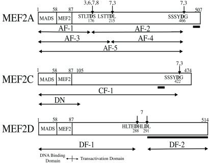
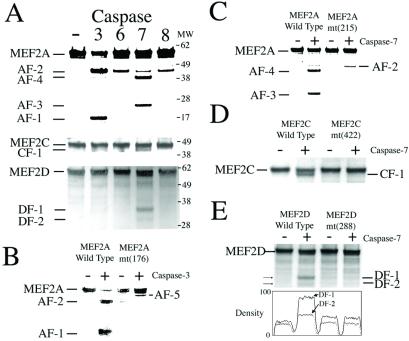
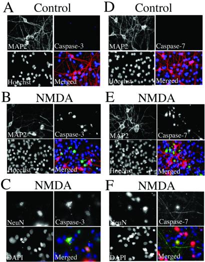
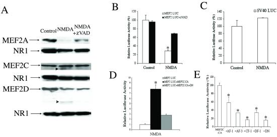
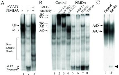
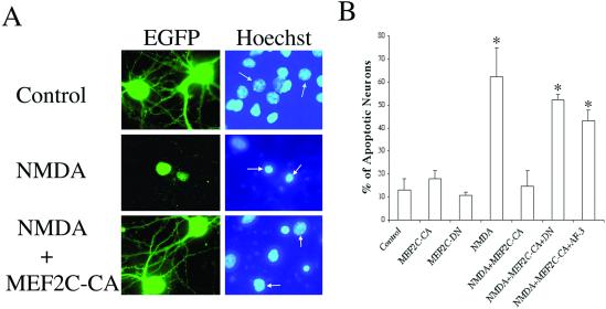
Myocyte enhancer factor-2 (MEF2) transcription factors are activated by p38 mitogen-activated protein kinase during neuronal and myogenic differentiation. Recent work has shown that stimulation of this pathway is antiapoptotic during development but proapoptotic in mature neurons exposed to excitotoxic or other stress. We now report that excitotoxic (N-methyl-D-aspartate) insults to mature cerebrocortical neurons activate caspase-3, -7, in turn cleaving MEF2A, C, and D isoforms. MEF2 cleavage fragments containing a truncated transactivation domain but preserved DNA-binding domain block MEF2 transcriptional activity via dominant interference. Transfection of constitutively active MEF2 (MEF2C-CA) rescues MEF2 transcriptional activity after N-methyl-D-aspartate insult and prevents neuronal apoptosis. Conversely, dominant-interfering MEF2 abrogates neuroprotection by MEF2C-CA. These results define a pathway to excitotoxic neuronal stress/apoptosis via caspase-catalyzed cleavage of MEF2. Additionally, we show that similar MEF2 cleavage fragments are generated in vivo during focal stroke damage. Hence, this pathway appears to have pathophysiological relevance in vivo.


Figures

Figure 1
Figure 1
Caspase cleavage sites of MEF2 proteins. MEF2 proteins contain a conserved N-terminal DNA-binding domain (with MADS and MEF2 domains; amino acids 1–87) and a transactivation domain. Observed caspase cleavage sites on MEF2 family proteins are indicated by arrows (caspases are listed in order of preference for substrate, as judged by density of bands in the autoradiograms of Fig. 2). The caspase-cleaved fragments are designated AF-1 to -5 for MEF2A, CF-1 for MEF2C, and DF-1 and -2 for MEF2D. The MEF2C-DN construct (DN) contains the first 105 amino acids (pcDNA1-MEF2C-1–105flag) and acts as a dominant negative because it lacks the full transactivation domain. The bar underneath the C-terminal segment of each transactivation domain indicates the region that is recognized by isoform-specific MEF2 antibodies.
Figure 2
Figure 2
Identification of caspase cleavage sites on MEF2 proteins. (A) In vitro translated, [35S]methionine-labeled MEF2A, -C, or -D proteins were incubated with purified caspase-3, -6, -7, or -8. Cleaved fragments (corresponding to those shown in Fig. 1) were resolved on NuPAGE and visualized by autoradiography. (B) Mutation of residue 176 of MEF2A from Asp to Ala [MEF2A mt(176)] prevented formation of AF-1 and -2 but enhanced AF-5 formation by caspase-3. (C) Mutation of residue 215 of MEF2A from Asp to Ala [MEF2A mt(215)] prevented formation of AF-3 and -4 by caspase-7. (D) Mutation of residue 422 of MEF2C from Asp to Ala [MEF2C mt(422)] prevented formation of CF-1 by caspase-7. (E) Mutation of residue 288 of MEF2D from Asp to Ala [MEF2D mt(288)] prevented formation of DF-1 and -2. This effect was quantified by densitometry.
Figure 3
Figure 3
Activation of neuronal caspase-3 and -7 after NMDA stimulation. Sham-treated (A and D) and NMDA-exposed (B, C, E, F) cerebrocortical cells were fixed and stained with Hoechst or 4′,6-diamidino-2-phenylindole (DAPI) nuclear dye (blue in merged images), anti-MAP-2 or anti-NeuN antibody (red), and antiactivated caspase-3 antibody (green in A–C) or antiactivated caspase-7 antibody (green in D–F). Stained preparations were examined under deconvolution microscopy. Note that images obtained 9 h after exposure to NMDA, a time when neurons destined to undergo apoptosis manifest caspase activity, have often lost their MAP-2 staining (29, 30). Hence, to confirm that these caspases were located specifically in neurons, we used a second neuronal marker more resistant to the initial phases of apoptosis, NeuN.
Figure 4
Figure 4
Caspase-mediated MEF2 cleavage inhibits MEF2 transcriptional activity after NMDA insult. (A) Western blots of cell lysates from control, NMDA-exposed, and NMDA plus zVAD.fmk-treated cultures probed with specific antibodies to MEF2A, -C, or -D. The blots were stripped and incubated with anti-NR1 antibody (the principal subunit of the NMDA receptor) as a loading control. Arrowheads indicate cleaved MEF2C or -D; these cleavage fragments correspond by molecular weight to CF-1 and DF-2, respectively (see Figs. 1 and 2). (B) NMDA insult reduces MEF2 transcriptional activity in luciferase reporter gene assays. The decrement in luciferase activity was prevented by zVAD.fmk (in other experiments, the control compound zFA.fmk had no effect). Cerebrocortical neurons were transfected with a MEF2 reporter gene (MEF2 LUC) before exposure to NMDA ± zVAD. Luciferase activity was measured 4 h after NMDA insult (NMDA-induced killing did not occur until 12–24 h). Baseline activity from untreated cells was assigned a value of 100% (values are mean ± SE; *, P < 0.001 by ANOVA). (C) As a control, transcriptional activity monitored in cells transfected with the simian virus 40 (SV40) enhancer-driven luciferase construct (SV40 LUC) was unaffected after NMDA exposure. (D) Constitutively active MEF2 (MEF2C-CA) prevented the decrease in MEF2 transcriptional activity engendered by NMDA exposure, but this effect was blocked by coexpression of a dominant-interfering form of MEF2 (MEF2C-DN, representing cleavage of MEF2 in the transactivation domain). (E) Truncated MEF2 proteins inhibit MEF2 transcriptional activity. Neurons were transfected with a MEF2 reporter gene (MEF2 LUC), MEF2C-CA, and expression vectors for the caspase cleavage fragments of MEF2 proteins (AF-1, AF-3, CF-1, DF-1) or dominant-negative MEF2 (DN). One day after transfection, luciferase activity was measured. In this case, activity observed after MEF2C-CA transfection was assigned a value of 100%.
Figure 5
Figure 5
EMSA for MEF2 full-length protein and cleavage fragment binding to the MEF2 site on DNA. (A) EMSA for MEF2. Cerebrocortical cultures were incubated for 2 h with vehicle (DMSO, lanes 1 and 2) or zVAD.fmk (lane 3) before stimulation with 100 μM NMDA for 20 min (lanes 2 and 3). Nuclear extracts were prepared 4 h after NMDA insult. EMSAs were performed by using 10 μg of nuclear cell extracts and 32P-labeled oligonucleotide representing the MEF2-binding site. Arrows: A/D, heteromer of full-length MEF2A and MEF2D binding to DNA (as determined by supershift assay; see below); A/C, heteromer of full-length MEF2A and MEF2C binding to DNA (as determined by supershift assay; see below). Arrowheads: low mobility complexes of MEF2 cleavage fragments and DNA. (B) Supershift analysis. Nuclear extracts were prepared from untreated (lanes 1–4) or NMDA exposed (lanes 5–8) cerebrocortical cultures. The nuclear lysates were preincubated with specific anti-MEF2 antibodies and then incubated with 32P-labeled oligonucleotide representing the MEF2-binding site. Arrows: SSC, supershifted complex of full-length MEF2 and DNA; A/D, heteromer of full-length MEF2A and MEF2D binding to DNA; A/C, heteromer of full-length MEF2A and MEF2C binding to DNA. Bottom of gel: low mobility complexes of MEF2 cleavage fragments and DNA. (C) MEF2 cleavage in vivo after stroke. Nuclear extracts were prepared 3 h after focal ischemia/reperfusion to one hemisphere or from the control undamaged hemisphere (n = 6 mice). Representative EMSA shows the presence of MEF2 cleavage fragments that bind to DNA only from the side with stroke damage (arrowhead).
Figure 6
Figure 6
Dependence of NMDA-induced neuronal apoptosis on MEF2 transcriptional activity. (A) NMDA-induced neuronal apoptosis was prevented by constitutively active MEF2. Cerebrocortical neurons were transfected with an empty vector or an expression vector encoding MEF2C-CA in conjunction with an EGFP expression vector to label transfected cells (green). One day after exposure to NMDA, apoptotic cells were scored by morphological changes with Hoechst nuclear staining (blue); neurons labeled by EGFP are marked by an arrow. Cells were verified to be neurons with MAP-2 staining (not shown). (B) Dominant-interfering forms of MEF2 prevent the neuroprotective effect of constitutively active MEF2. Neurons were transfected with an EGFP expression vector plus expression vectors for control (empty vector), MEF2C-CA, MEF2C-DN, or a caspase-cleaved form of MEF2A (AF-3). Apoptosis in transfected neurons was as-sessed with Hoechst nuclear staining. MEF2C-CA ameliorated NMDA-induced neuronal apoptosis, whereas dominant-interfering forms of MEF2 (MEF2-DN and AF-3) blocked this neuroprotective effect. Values are mean ± SE; *, P < 0.001 by ANOVA.

References

    1. Kawasaki H, Morooka T, Shimohama T, Gotoh Y, Nishida E. J Biol Chem. 1997;272:18518–18521. - PubMed
    1. Kikuchi M, Tenneti L, Lipton S A. J Neurosci. 2000;20:5037–5044. - PMC - PubMed
    1. Mukherjee P K, DeCoster M A, Campbell F Z, Davis R J, Bazan N G. J Biol Chem. 1999;274:6493–6498. - PubMed
    1. Okamoto S-i, Krainc D, Sherman K, Lipton S A. Proc Natl Acad Sci USA. 2000;97:7561–7566. - PMC - PubMed
    1. Han J, Jiang Y, Li Z, Kravchenko V V, Ulevitch R J. Nature (London) 1997;386:296–299. - PubMed

Publication types

MeSH terms