

Increased expression of the neuronal glutamate transporter (EAAT3/EAAC1) in hippocampal and neocortical epilepsy
- PMID: 11906504
- PMCID: PMC2441873
- DOI: 10.1046/j.1528-1157.2002.35001.x
Increased expression of the neuronal glutamate transporter (EAAT3/EAAC1) in hippocampal and neocortical epilepsy
Abstract
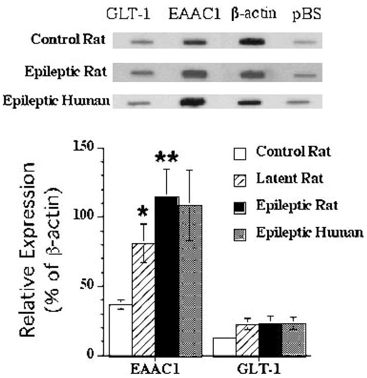
Purpose: To define the changes in gene and protein expression of the neuronal glutamate transporter (EAAT3/EAAC1) in a rat model of temporal lobe epilepsy as well as in human hippocampal and neocortical epilepsy.
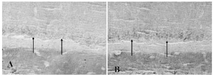
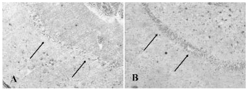
Methods: The expression of EAAT3/EAAC1 mRNA was measured by reverse Northern blotting in single dissociated hippocampal dentate granule cells from rats with pilocarpine-induced temporal lobe epilepsy (TLE) and age-matched controls, in dentate granule cells from hippocampal surgical specimens from patients with TLE, and in dysplastic neurons microdissected from human focal cortical dysplasia specimens. Immunolabeling of rat and human hippocampi and cortical dysplasia tissue with EAAT3/EAAC1 antibodies served to corroborate the mRNA expression analysis.
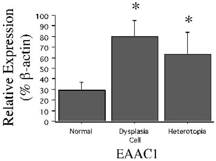
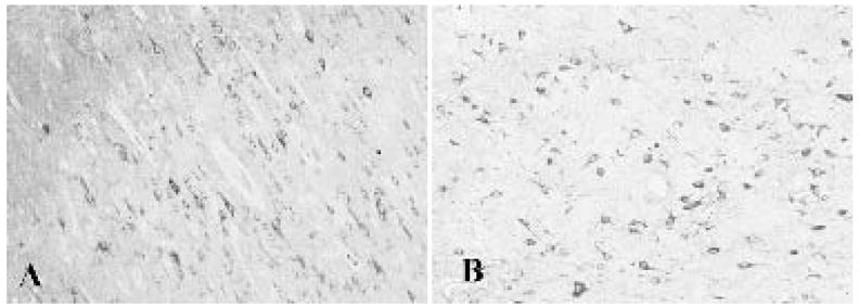
Results: The expression of EAAT3/EAAC1 mRNA was increased by nearly threefold in dentate granule cells from rats with spontaneous seizures compared with dentate granule cells from control rats. EAAT3/EAAC1 mRNA levels also were high in human dentate granule cells from patients with TLE and were significantly elevated in dysplastic neurons in cortical dysplasia compared with non-dysplastic neurons from postmortem control tissue. No difference in expression of another glutamate transporter, EAAT2/GLT-1, was observed. Immunolabeling demonstrated that EAAT3/EAAC1 protein expression was enhanced in dentate granule cells from both rats and humans with TLE as well as in dysplastic neurons from human cortical dysplasia tissue.
Conclusions: Elevations of EAAT3/EAAC1 mRNA and protein levels are present in neurons from hippocampus and neocortex in both rats and humans with epilepsy. Upregulation of EAAT3/EAAC1 in hippocampal and neocortical epilepsy may be an important modulator of extracellular glutamate concentrations and may occur as a response to recurrent seizures in these cell types.
Figures
References
-
- Greenamyre JT. The role of glutamate in neurotransmission and in neurologic disease. Arch Neurol. 1986;43:1058–63. - PubMed
-
- Choi DW. Excitotoxic cell death. J Neurobiol. 1992;23:1261–76. - PubMed
-
- Sims KD, Robinson MB. Expression patterns and regulation of glutamate transporters in the developing and adult nervous system. Crit Rev Neurobiol. 1999;13:169–97. - PubMed
-
- Dabolt NC. Glutamate uptake. Prog Neurobiol. 2001;65:1–105. - PubMed
-
- Robinson MB, Dowd LA. Heterogeneity and functional properties of subtypes of sodium-dependent glutamate transporters in the mammalian central nervous system. Adv Pharmacol. 1997;37:69–115. - PubMed
Publication types
MeSH terms
Substances
Grants and funding
- NS38572/NS/NINDS NIH HHS/United States
- R01 NS036465/NS/NINDS NIH HHS/United States
- NS36465/NS/NINDS NIH HHS/United States
- MH01658/MH/NIMH NIH HHS/United States
- NS38595/NS/NINDS NIH HHS/United States
- R01 NS029868/NS/NINDS NIH HHS/United States
- NS39938/NS/NINDS NIH HHS/United States
- R01 NS038572/NS/NINDS NIH HHS/United States
- R01 NS038595/NS/NINDS NIH HHS/United States
- NS32403/NS/NINDS NIH HHS/United States
- NS29868/NS/NINDS NIH HHS/United States
- R01 NS032403/NS/NINDS NIH HHS/United States
- R37 NS032403/NS/NINDS NIH HHS/United States
LinkOut - more resources
Full Text Sources
