. 2002 May 24;277(21):18868-74.
doi: 10.1074/jbc.M201463200. Epub 2002 Mar 20.

Biochemical and structural definition of the l-afadin- and actin-binding sites of alpha-catenin

Affiliations

Biochemical and structural definition of the l-afadin- and actin-binding sites of alpha-catenin

Sabine Pokutta et al. J Biol Chem. .

Abstract

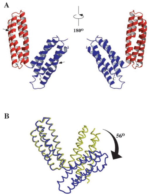
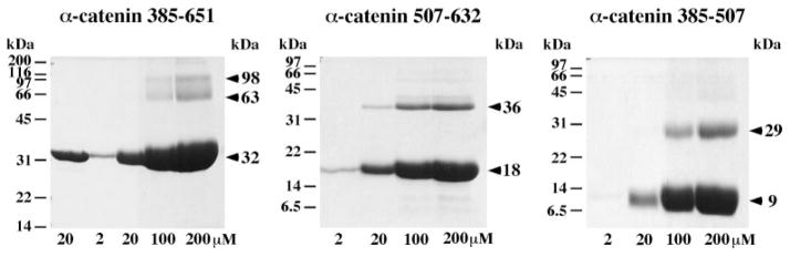
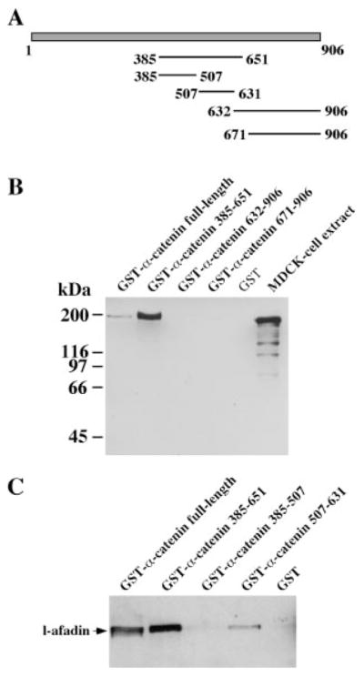
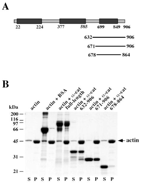
alpha-Catenin is an integral component of adherens junctions, where it links cadherins to the actin cytoskeleton. alpha-Catenin is also required for the colocalization of the nectin/afadin/ponsin adhesion system to adherens junctions, and it specifically associates with the nectin-binding protein afadin. A proteolytic fragment of alpha-catenin, residues 385-651, contains the afadin-binding site. The three-dimensional structure of this fragment comprises two side-by-side four-helix bundles, both of which are required for afadin binding. The alpha-catenin fragment 385-651 binds afadin more strongly than the full-length protein, suggesting that the full-length protein harbors a cryptic binding site for afadin. Comparison of the alpha-catenin 385-651 structure with the recently solved structure of the alpha-catenin M-fragment (Yang, J., Dokurno, P., Tonks, N. K., and Barford, D. (2001) EMBO J. 20, 3645-3656) reveals a surprising flexibility in the orientation of the two four-helix bundles. alpha-Catenin and the actin-binding protein vinculin share sequence and most likely structural similarity within their actin-binding domains. Despite this homology, actin binding requires additional sequences adjacent to this region.


Figures

Fig. 1
Fig. 1. Structure of α-catenin 385–651
A, ribbon diagram of the structure; the two views of the molecule rotated by 180°. The N- and C-terminal four-helix bundles are colored in red and blue, respectively. These subdomains correspond to the N- and C-terminal fragments used in cross-linking and afadin binding studies. The positions of proline residues within the helices are indicated by an arrow. B, relative motion of the N- and C-terminal subdomains of α-catenin 385–651. The N-terminal subdomains of the α-catenin 385–651 fragment (yellow) and the α-catenin M fragment (Ref. ; residues 377–633) (blue) were superimposed. The relative orientation of the N- and C-terminal domain varies in the five different views obtained from the two different crystal forms. The two copies shown here show the largest angular displacement between the two four-helix bundles. The figure was prepared using MOLSCRIPT (39) and RASTER3D (40).
Fig. 2
Fig. 2. Oligomerization of α-catenin 385–651 and its N- and C-terminal subdomains
Fragments at the indicated concentrations were incubated with a 30-fold excess of cross-linker. For the two amine-reactive cross-linking reagents BS3 and DMS with spacer arms of 11.4 and 11.0 Å, respectively, different cross-linking efficiency was observed. Higher cross-linking efficiency results are shown here and were obtained with DMS in the case of α-catenin 507–632 and BS3 for α-catenin 385–651 and α-catenin 385–507. The first lane for the α-catenin 385–651 fragment shows a sample in the absence of cross-linker. Molecular mass markers are indicated on the left of each gel, and apparent molecular mass of the fragments and cross-linking products are shown on the right.
Fig. 3
Fig. 3. Interaction of α-catenin with l-afadin
A, schematic representation of the different α-catenin fragments used in the binding assays. B, proteins expressed as GST fusion proteins were coupled to glutathione-agarose beads and incubated with MDCK cell lysate. Bound protein was analyzed by Western blotting using monoclonal anti-afadin antibody. The signal for l-afadin in 15 μl of MDCK lysate, which corresponds to 1% of the amount used in the binding assays, is shown in the right lane. Molecular mass markers are shown on the left. C, binding to the N- and C-terminal subdomains of α-catenin 385–651.
Fig. 4
Fig. 4. Interaction of α-catenin (α-cat) with F-actin
A, schematic representation of α-catenin and the constructs used in the binding assay. Vinculin homology regions are shown in dark gray. B, binding to F-actin was examined by cosedimentation. Assuming a binding ratio of protein:monomeric actin of 1:7, all α-catenin constructs were added in excess. The supernatant (S) containing the unbound protein and the pellet (P) containing F-actin and bound protein are shown for each sample. Molecular mass markers are indicated on the left.

References

    1. Gumbiner BM. Cell. 1996;84:345–357. - PubMed
    1. Tomschy A, Fauser C, Landwehr R, Engel J. EMBO J. 1996;15:3507–3514. - PMC - PubMed
    1. Brieher WM, Yap AS, Gumbiner BM. J Cell Biol. 1996;135:487–496. - PMC - PubMed
    1. Steinberg MS, McNutt PM. Curr Opin Cell Biol. 1999;11:554–560. - PubMed
    1. Adams CL, Nelson WJ, Smith SJ. J Cell Biol. 1996;135:1899–1911. - PMC - PubMed

Publication types

Associated data

LinkOut - more resources