. 2002 Apr;76(8):3709-19.
doi: 10.1128/jvi.76.8.3709-3719.2002.

Parameters of human hepatitis delta virus genome replication: the quantity, quality, and intracellular distribution of viral proteins and RNA

Affiliations

Parameters of human hepatitis delta virus genome replication: the quantity, quality, and intracellular distribution of viral proteins and RNA

Severin Gudima et al. J Virol. 2002 Apr.

Abstract

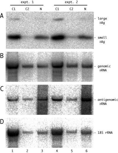
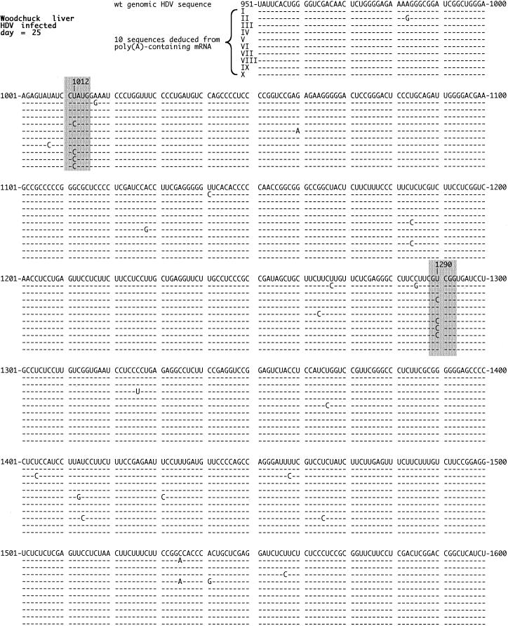
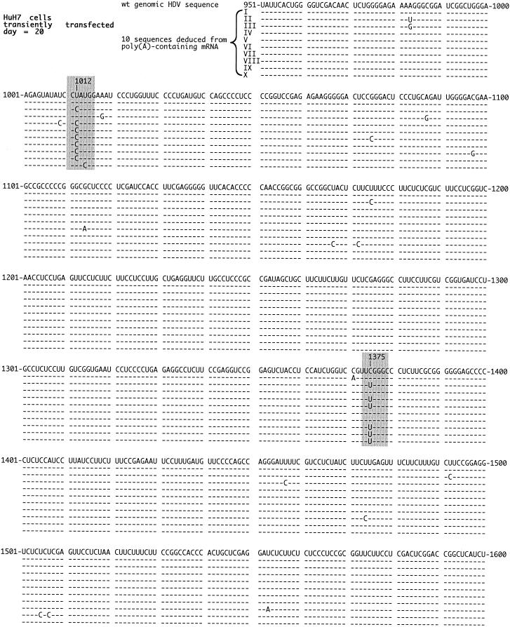
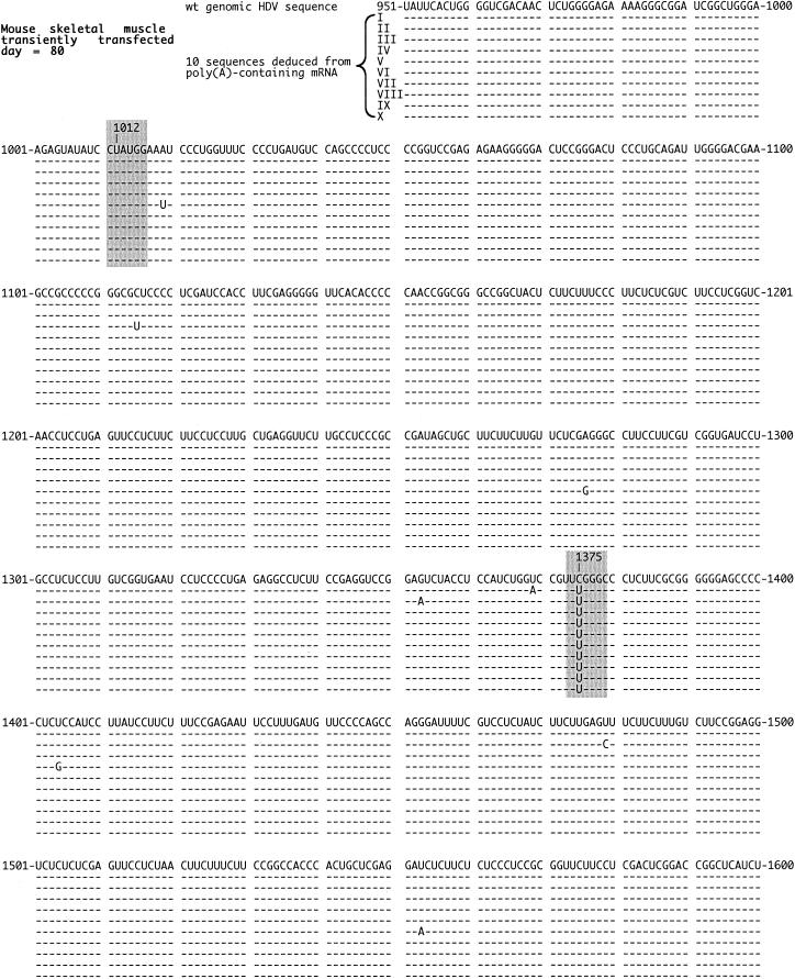
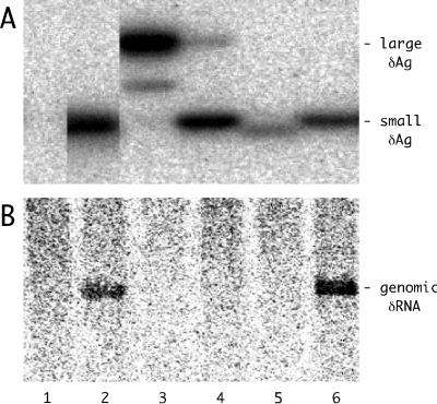
Assembly of hepatitis delta virus (HDV) in infected human hepatocytes involves association of the 1,679- nucleotide single-stranded genomic RNA (deltaRNA) with multiple copies of both small and large forms of the delta protein (deltaAg) to form a ribonucleoprotein particle which in turn interacts with envelope proteins of the natural helper virus, hepatitis B virus. Subsequently, for initiation of a new round of replication, the amount of small deltaAg within the assembled HDV particle is both necessary and sufficient. Quantitative assays were used in order to better understand just how much deltaAg is needed. The molar ratio of deltaAg species to genomic deltaRNA in assembled HDV particles was approximately 200. Next, this ratio was determined for cells under several different experimental situations in which HDV genome replication was occurring. These included replication in woodchuck liver and also in mouse liver and skeletal muscle, as well as replication in stably and transiently transfected cultured human hepatoblastoma cells. Surprisingly, in almost all these situations the molar ratios were comparable to that observed for HDV particles. This was true for different times after the initiation of replication and was independent of whether or not virus assembly was occurring. Cell fractionation combined with quantitative assays was used to test whether the majority of deltaAg and deltaRNA were colocalized during HDV replication in transfected cells. The cytoplasmic fraction contained the majority of deltaAg and genomic deltaRNA. Finally, the quality of deltaAg and deltaRNA, especially at relatively late times after the initiation of replication, was examined by using reverse transcription-PCR, cloning, and sequencing through the entire deltaAg open reading frame. When virus assembly and spread were not possible, 20% or less of the predicted deltaAg would have been able to support HDV replication. In summary, an examination of the quantity, quality and intracellular distribution of deltaAg and deltaRNA in several different experimental systems has provided a better understanding of the parameters associated with the initiation, maintenance, and ultimate decline of HDV genome replication.


Figures

FIG. 1.
FIG. 1.
Detection of δAg and genomic and antigenomic δRNA in fractions of HuH7 cells transfected with HDV RNAs. Duplicate cultures were transfected with antigenomic HDV RNAs. After 12 days the cells were fractionated to provide the cytoplasmic fractions C1 and C2 and the final nuclear fraction N. The total protein and RNA were extracted from each of these fractions. Panels A to D show the results of assays for δAg, genomic δRNA, antigenomic δRNA, and 18S rRNA, respectively. Note that in panel A we detected both small and large forms of δAg.
FIG. 2.
FIG. 2.
Quality of HDV RNA detected in infected woodchuck liver. At the peak of an acute HDV infection of a woodchuck, the animal was sacrificed and total RNA was extracted from the liver. The poly(A)-containing RNA was then isolated, and the region spanning the ORF for the δAg was subjected to RT-PCR, cloning, and sequencing. The figure shows 10 sequences deduced for genomic RNA in relationship to the small δAg wild-type sequence of Kuo et al., using the same numbering system (21). Highlighted in gray are the changes at nt 1012 and 1290 that occurred multiple times and are discussed in the text. The nonhighlighted changes did not occur more than twice. Note that the 3 nt changes on sequences III and VI, in addition to that at nt 1290, were silent in terms of the predicted amino acid sequence.
FIG. 3.
FIG. 3.
Quality of HDV RNA detected in RNA-transfected HuH7 cells. Total RNA was extracted from HuH7 cells at 20 days after RNA transfection. Otherwise, details are as described in the legend to Fig. 2. Highlighted in gray are the changes at nt 1012 and 1375. An 11th sequence, not shown, was identical to that of the wild type.
FIG. 4.
FIG. 4.
Quality of HDV RNA detected in DNA-transfected mouse liver. Total RNA was extracted from mouse liver at day 30 after DNA transfection. Otherwise, details are as described in the legend to Fig. 2. Highlighted in gray are the changes at nt 1012 and 1375. An 11th sequence, not shown, was identical to that of the wild type.
FIG. 5.
FIG. 5.
Quality of HDV RNA detected in DNA-transfected mouse skeletal muscle. Total RNA was extracted from mouse skeletal muscle at day 80 after DNA transfection. Otherwise, details are as described in the legend to Fig. 2. As highlighted in gray, no changes were detected at nt 1012, while 10 of 10 sequences were changed at nt 1375.
FIG. 6.
FIG. 6.
Expression and functional tests of the R(75)Q small δAg produced following the change at nt 1375. In lanes 1 to 6, HuH7 cells were each transfected with pDL481, a plasmid expressing greater-than-unit-length antigenomic HDV RNA with a frameshift mutation in the δAg ORF (20, 43), along with the indicated combinations of plasmids expressing the small, large, or mutated small forms of δAg. The transfection strategy was as previously described (7). The expression of the mutated small form of δAg was from pSG206, constructed by modification of pDL444, the plasmid expressing wild-type small δAg. In lanes 2 to 6, the cotransfection included the following: lane 2, plasmids expressing wild-type small δAg; lane 3, wild-type large δAg; lane 4, both small and large δAg in the ratio 0.8:0.2; lane 5, mutant δAg; lane 6, mutant δAg and small δAg in the ratio 0.5:0.5. Panels A and B show the immunoblot and Northern analyses, respectively.

References

    1. Ausubel, F. M., R. Brent, R. E. Kingston, D. D. Moore, J. A. Smith, J. G. Seidman, and K. Struhl. 1987. Current protocols in molecular biology. Wiley, New York, N.Y.
    1. Bell, P., R. Brazas, D. Ganem, and G. G. Maul. 2000. Hepatitis delta virus replication generates complexes of large hepatitis delta antigen and antigenomic RNA that affiliate with and alter nuclear domain 10. J. Virol. 74:5329-5336. - PMC - PubMed
    1. Bichko, V. V., and J. M. Taylor. 1996. Redistribution of the delta antigens in cells replicating the genome of hepatitis delta virus. J. Virol. 70:8064-8070. - PMC - PubMed
    1. Casey, J. L., and J. L. Gerin. 1995. Hepatitis D virus RNA editing: specific modification of adenosine in the antigenomic RNA. J. Virol. 69:7593-7700. - PMC - PubMed
    1. Chang, F. L., P. J. Chen, S. J. Tu, M. N. Chiu, C. J. Wang, and D. S. Chen. 1991. The large form of hepatitis δ antigen is crucial for the assembly of hepatitis δ virus. Proc. Natl. Acad. Sci. USA 88:8490-8494. - PMC - PubMed

Publication types

MeSH terms

LinkOut - more resources