. 2002 Apr;76(8):3739-47.
doi: 10.1128/jvi.76.8.3739-3747.2002.

Human immunodeficiency virus cDNA metabolism: notable stability of two-long terminal repeat circles

Affiliations

Human immunodeficiency virus cDNA metabolism: notable stability of two-long terminal repeat circles

Scott L Butler et al. J Virol. 2002 Apr.

Abstract

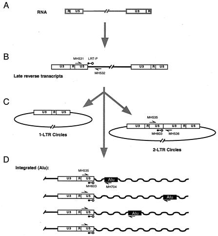
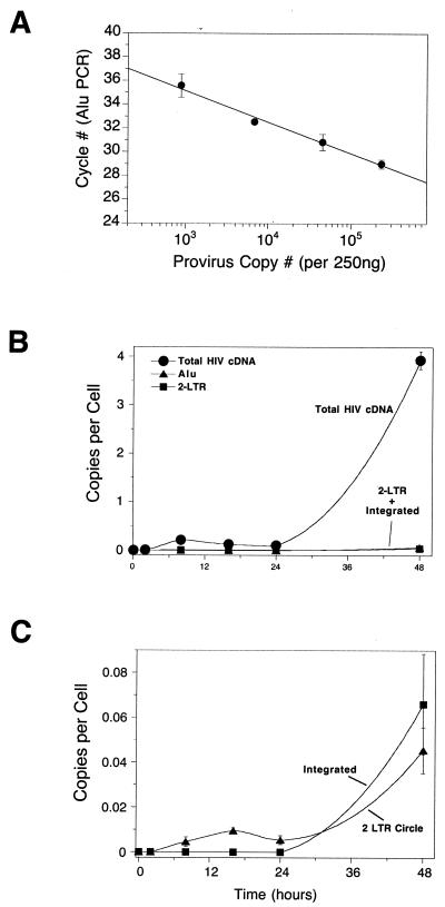
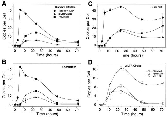
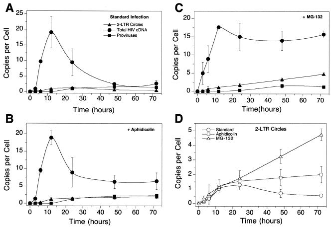
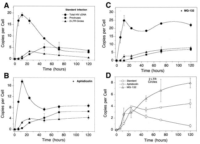
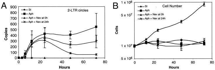
Early steps of retroviral replication involve reverse transcription of the viral RNA to yield a linear double-stranded cDNA copy and then integration of the viral cDNA into a chromosome of the host cell. A portion of the viral cDNA can also follow nonproductive pathways in which it becomes circularized. In one pathway, the ends of the linear cDNA become joined together by the cellular nonhomologous DNA end-joining system to form two-long terminal repeat (2-LTR) circles. It has been argued that 2-LTR circles are quickly degraded in human immunodeficiency virus (HIV)-infected cells, allowing the presence of 2-LTR circles to be used as a marker for ongoing de novo infection in patients. Following this idea, detection of 2-LTR circles in patients undergoing successful highly active antiretroviral therapy has led to the proposal that viral replication persists despite treatment. We have used fluorescence-monitored PCR (Taqman) to quantitate the metabolism of HIV cDNA early after infection. Contrary to previous work, we find that 2-LTR circles are actually quite stable in experiments where confounding variables are controlled. Thus, studies relying on the lability of 2-LTR circles are open to reinterpretation. We also used the quantitative PCR methods to analyze the effects of MG132, a proteasome inhibitor, which revealed that viral complexes containing mostly completed cDNAs are the primary substrates for proteasome-mediated degradation.


Figures

FIG. 2.
FIG. 2.
HIV cDNA metabolism in a multicycle HIV infection of SupT1 cells assayed by quantitative PCR. (A) Standard curve relating the number of integrated HIV cDNA copies quantitated by the total cDNA amplicon to the Alu PCR cycle of threshold. Using the standard curve the number of proviruses can be determined from a measurement of the Alu PCR signal. (B) Accumulation of total HIV cDNA (i.e., cDNAs that have completed the second strand transfer step of reverse transcription), 2-LTR circles, and integrated proviruses during multicycle HIV infection. In this range of copies per cell (y axis) the 2-LTR circles and integrated provirus signals are at the baseline. In this and subsequent figures, total HIV cDNA copies are indicated by filled circles, 2-LTR circles are indicated by filled triangles, and integrate proviruses are indicated by filled squares. (C) Accumulation of 2-LTR circles and integrated proviruses replotted against lower levels of copies per cell (y axis). The multiplicity of infection was 0.04 infectious unit per cell as determined by infection of 5.25 indicator cells. Error bars, standard deviations.
FIG. 3.
FIG. 3.
HIV cDNA metabolism in single-cycle infections of SupT1 cells with HIV-1 assayed by quantitative PCR. (A) Infection of SupT1 cells with HIV R9 in the absence of added cell cycle inhibitors. The numbers of copies of each cDNA form per cell are as indicated. Points are labeled as in Fig. 1. (B) Infection as shown in panel A but with aphidicolin (10 μg/ml). (C) Infection as shown in panel A but with 50 μM MG132. (D) Comparison of 2-LTR circle formation in the assays in panels A to C. Error bars, standard deviations.
FIG. 1.
FIG. 1.
Diagram of the early steps of HIV infection, indicating the locations of primers used for the quantitative PCR assays. The RNA genome (A) is converted to a linear double-stranded cDNA form by reverse transcription (B). Some of the viral cDNA is circularized (C), while other HIV cDNA molecules are integrated in to the cellular DNA (D). Different proviruses are integrated at different distances from cellular Alu repeats.
FIG. 4.
FIG. 4.
Metabolism of cDNA synthesized by infection with the SM2 HIV-based vector. (A) Infection of SupT1 cells with the SM2 vector in the absence of added cell cycle inhibitors. (B) Infection as shown in panel A but with aphidicolin (10 μg/ml). (C) Infection as shown in panel A but with 50 μM MG132. (D) Comparison of 2-LTR circle formation in the assays in panels A to C. Error bars, standard deviations.
FIG. 5.
FIG. 5.
Metabolism of cDNA synthesized by infection with the SIN HIV-based vector. (A) Infection of SupT1 cells with the SIN vector in the absence of added cell cycle inhibitors. (B) Infection as shown in panel A but with aphidicolin (10 μg/ml). (C) Infection as shown in panel A but with 50 μM MG132. (D) Comparison of 2-LTR circle formation in the assays in panels A to C. Error bars, standard deviations.
FIG. 6.
FIG. 6.
A covert steady-state model does not account for the observed stability of 2-LTR circles. Cells were infected with the SM2 HIV-based vector (spinoculation was not used), and aliquots were analyzed. Cells were treated with 0.3 μM aphidicolin (Aph), aphidicolin plus nevirapine at time zero (Aph + Nev at 0 h), aphidicolin plus nevirapine at 24 h (Aph + Nev at 24 h), or no added drug (St [for “standard”]). (A) Assay of 2-LTR circle DNA. (B) Viable cell number in cultures studied (assayed by trypan blue exclusion), indicating that aphidicolin arrested the cell cycle. Error bars, standard deviations.

References

    1. Bell, P., L. J. Montaner, and G. G. Maul. 2001. Accumulation and intranuclear distribution of unintegrated human immunodeficiency virus type 1 DNA. J. Virol. 75:7683-7691. - PMC - PubMed
    1. Butler, S., M. Hansen, and F. D. Bushman. 2001. A quantitative assay for HIV cDNA integration in vivo. Nat. Med. 7:631-634. - PubMed
    1. Coffin, J. M., S. H. Hughes, and H. E. Varmus. 1997. Retroviruses. Cold Spring Harbor Laboratory Press, Cold Spring Harbor, N.Y. - PubMed
    1. Follenzi, A., L. E. Ailes, S. Bakovic, M. Gueuna, and L. Naldini. 2000. Gene transfer by lentiviral vectors is limited by nuclear translocation and rescued by HIV-1 pol sequences. Nat. Genet. 25:217-222. - PubMed
    1. Hansen, M. S. T., G. J. I. Smith, T. Kafri, V. Molteni, J. S. Siegel, and F. D. Bushman. 1999. Integration complexes derived from HIV vectors for rapid assays in vitro. Nat. Biotechnol. 17:578-582. - PubMed

Publication types

LinkOut - more resources