. 2002 Apr;76(8):3756-64.
doi: 10.1128/jvi.76.8.3756-3764.2002.

Membrane recognition by vesicular stomatitis virus involves enthalpy-driven protein-lipid interactions

Affiliations

Membrane recognition by vesicular stomatitis virus involves enthalpy-driven protein-lipid interactions

Fabiana A Carneiro et al. J Virol. 2002 Apr.

Abstract

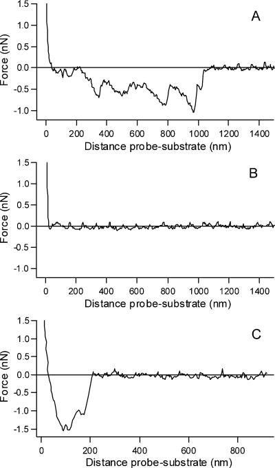
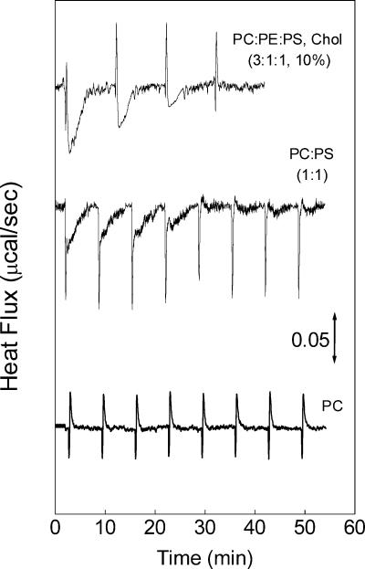
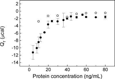
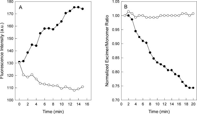
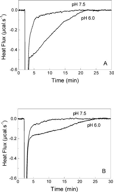
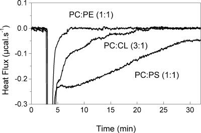
Vesicular stomatitis virus (VSV) infection depends on the fusion of viral and cellular membranes, which is mediated by virus spike glycoprotein G at the acidic environment of the endosomal compartment. VSV G protein does not contain a hydrophobic amino acid sequence similar to the fusion peptides found among other viral glycoproteins, suggesting that membrane recognition occurs through an alternative mechanism. Here we studied the interaction between VSV G protein and liposomes of different phospholipid composition by force spectroscopy, isothermal titration calorimetry (ITC), and fluorescence spectroscopy. Force spectroscopy experiments revealed the requirement for negatively charged phospholipids for VSV binding to membranes, suggesting that this interaction is electrostatic in nature. In addition, ITC experiments showed that VSV binding to liposomes is an enthalpically driven process. Fluorescence data also showed the lack of VSV interaction with the vesicles as well as inhibition of VSV-induced membrane fusion at high ionic strength. Intrinsic fluorescence measurements showed that the extent of G protein conformational changes depends on the presence of phosphatidylserine (PS) on the target membrane. Although the increase in PS content did not change the binding profile, the rate of the fusion reaction was remarkably increased when the PS content was increased from 25 to 75%. On the basis of these data, we suggest that G protein binding to the target membrane essentially depends on electrostatic interactions, probably between positive charges on the protein surface and negatively charged phospholipids in the cellular membrane. In addition, the fusion is exothermic, indicating no entropic constraints to this process.


Figures

FIG. 1.
FIG. 1.
Force-distance curves for VSV interaction with membranes. Force-distance curves were recorded on lipid-covered mica substrates. Retracting curves were obtained with VSV adsorbed on the tip and mica substrates covered with PC-PS (3:1) (A), PC only (B), or PC-CL (3:1) (C). The negative values for the force peaks in panels A and C indicate adhesion and are absent in panel B. Data were collected in 20 mM MES-30 mM Tris, pH 7.5, at room temperature.
FIG. 2.
FIG. 2.
Calorimetric measurement of VSV binding to liposomes at 35oC. Typical calorimetric traces (heat flow as a function of time) obtained for four to eight injections (5 μl each) of a VSV suspension (28 μg/ml) into the cell containing unilamellar vesicles of PC-PE-PS with cholesterol (3:1:1 and 10%), PC-PS (1:3), or PC only, in 20 mM MES-30 mM Tris, pH 7.5, at 35°C. The sharp peaks are due to the VSV dilution, as seen in control experiments of the injection of virus into buffer (not shown). The phospholipid concentration was 1 mM.
FIG. 3.
FIG. 3.
Binding isotherms. The total heat (QT) was calculated for each peak of the calorimetric thermograms resulting from the injection of VSV into vesicles (see Fig. 1). QT is plotted as a function of the protein concentration in each injection, with the mean ± standard error (SE) for five different experiments with PC-PS (•) and the mean of two experiments with PC-only vesicles (○) obtained with the same VSV preparation. The data were essentially the same for the PC-PS vesicles containing 25, 50, or 75% PS. The conditions were the same as in Fig. 1. Bar, 0.05 μcal s−1.
FIG. 4.
FIG. 4.
VSV G protein conformational change during virus incubation with vesicles of different phospholipid compositions. (A) Intrinsic fluorescence of VSV was recorded after virus incubation with small unilamellar vesicles of PC-PS (1:3) (•), PC-PS (1:1) (▴), PC-PS (3:1) (▪), and PC only (○). The vesicles were prepared in 20 mM MES-30 mM Tris buffer, pH 6.0, in a final phospholipid concentration of 0.1 mM. The excitation wavelength was 280 nm, and the emission was collected at 334 nm. The final protein concentration was 70 μg/ml. (B) Purified virus was added to a sample containing equal amounts of unlabeled vesicles and vesicles labeled with 10-PyPC. VSV-induced membrane fusion was measured by the decrease in the 10-PyPC excimer/monomer fluorescence intensity ratio. Vesicles used were PC-PS (1:3) (•), PC-PS (1:1) (▴), and PC-PS (3:1) (▪) at pH 6.0 and PC-PS (1:3) at pH 7.5 (○). 10-PyPC was excited at 340 nm, and the intensities were collected at 480 and 376 nm for the excimer and monomer, respectively. Experimental conditions were the same as described in the legend to panel A.
FIG. 5.
FIG. 5.
Effect of high ionic strength on VSV G protein conformational changes during interaction with liposomes. (A) Intrinsic fluorescence of VSV was recorded after virus incubation with vesicles composed of PC-PS (1:3) in the absence (•) and in the presence (○) of 250 mM KCl. The vesicles were prepared in 20 mM MES-30 mM Tris buffer, pH 6.0, in a final phospholipid concentration of 0.1 mM. The excitation wavelength was 280 nm, and the emission was collected at 334 nm. The final protein concentration was 70 μg/ml. (B) VSV-induced membrane fusion measured as in Fig. 4 after virus incubation with vesicles composed of PC-PS (1:3) in the absence (•) and in the presence (○) of 250 mM KCl. Experimental conditions were the same as described in the legend to panel A.
FIG. 6.
FIG. 6.
Calorimetric traces of the fusion of VSV with vesicles of different PS content at 35oC . The calorimetric traces were obtained after the injection of 10 μl of VSV solution (0.28 mg/ml) into the cell containing 1 mM vesicles of PC-PS (1:3) (A) and PC-PS (3:1) (B) at pH 6.0 or pH 7.5, as indicated in each panel. After the heat due to the VSV dilution, there is a negative heat effect that can be associated with the fusion process. The return to the baseline level indicates that the fusion was complete. At pH 7.5, only the heat effect associated with the VSV dilution and binding to the vesicles is observed. The samples were prepared in 20 mM MES-30 mM Tris buffer, pH 7.5 or 6.0.
FIG. 7.
FIG. 7.
Calorimetric traces of the fusion of VSV with vesicles of different PS content at 35oC . The calorimetric traces were obtained after the injection of 10 μl of VSV solution (0.28 mg/ml) into the cell containing 1 mM vesicles of PC-PE (1:1), PC-CL (3:1), and PC-PS (1:3). The vesicles were prepared in 20 mM MES-30 mM Tris buffer, pH 6.0.
FIG. 8.
FIG. 8.
Kinetics of VSV fusion with membranes at 35oC . The heat released after injection of VSV into the cell containing vesicles at pH 6.0 (•, ▪) or at pH 7.5 (○, □) was calculated by integrating the calorimetric traces shown in Fig. 6 for PC-PS at ratios of 1:3 (•, ○) and 3:1 (▪, □). Kinetics of the heat effects after VSV injection into vesicles of PC only at pH 6.0 (▴) was obtained from thermograms similar to those in Fig. 6 (not shown).

References

    1. Bailey, C. A., D. K. Miller, and J. Lenard. 1984. Effects of DEAE-dextran on infection and hemolysis by VSV. Evidence that nonspecific electrostatic interactions mediate effective binding of VSV to cells. Virology 133:111-117. - PubMed
    1. Baranowski, E., C. M. Ruiz-Jarabo, and E. Domingo. 2001. Evolution of cell recognition by viruses. Science 292:1102-1105. - PubMed
    1. Bathaie, S. Z., A. A. Moosavi-Movahedi, and A. A. Saboury. 1999. Energetic and binding properties of DNA upon interaction with dodecyl trimethylammonium bromide. Nucleic Acids Res. 27:1001-1005. - PMC - PubMed
    1. Bonnafous, P., and T. Stegmann. 2000. Membrane perturbation and fusion pore formation in influenza hemagglutinin-mediated membrane fusion. A new model for fusion. J. Biol. Chem. 275:6160-6166. - PubMed
    1. Bullough, P. A., F. M. Hughson, J. J. Skehel, and D. C. Wiley. 1994. Structure of influenza haemagglutinin at the pH of membrane fusion. Nature 371:37-43. - PubMed

Publication types

LinkOut - more resources