. 2002 Apr;76(8):4008-21.
doi: 10.1128/jvi.76.8.4008-4021.2002.

Persistent and transient replication of full-length hepatitis C virus genomes in cell culture

Affiliations

Persistent and transient replication of full-length hepatitis C virus genomes in cell culture

Thomas Pietschmann et al. J Virol. 2002 Apr.

Abstract

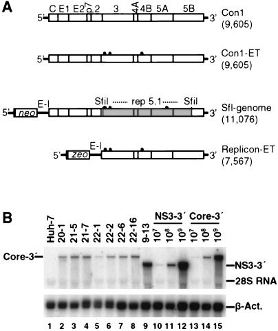
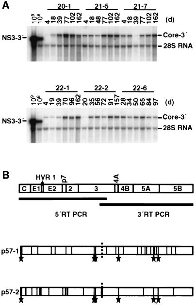
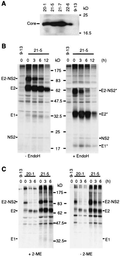
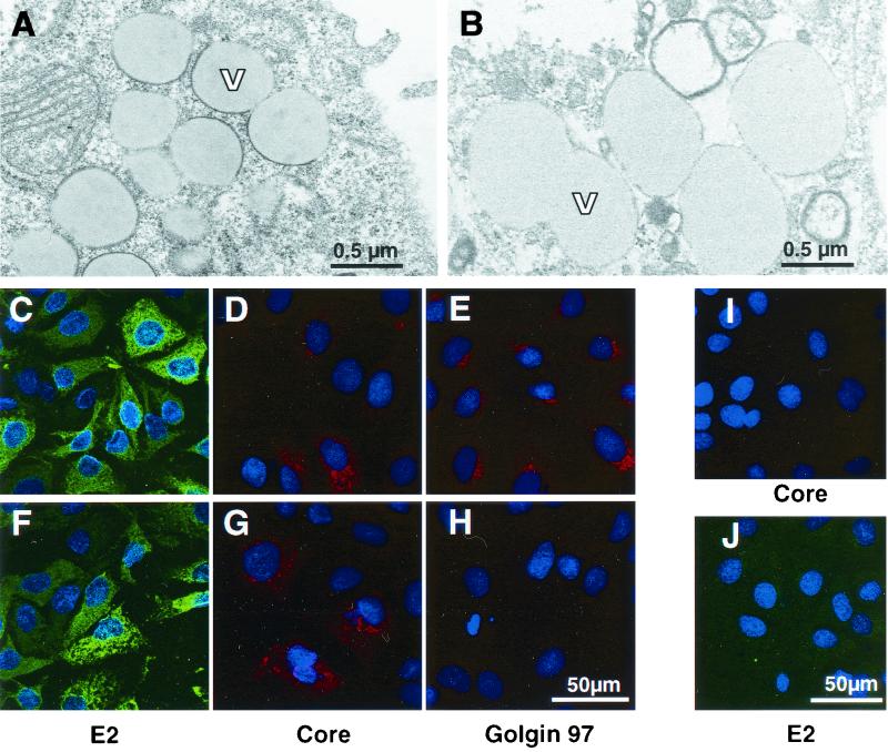
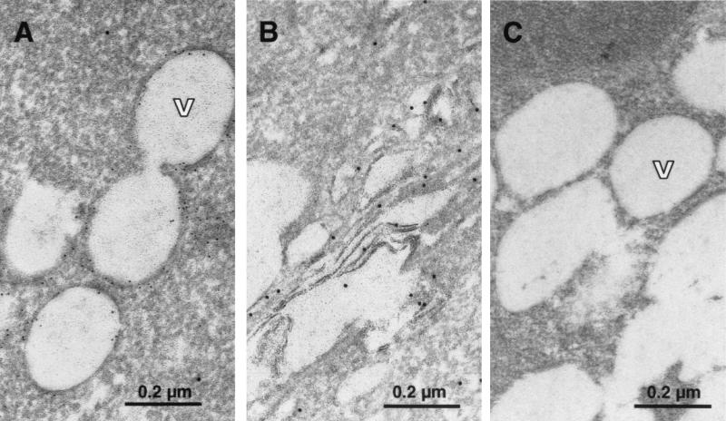
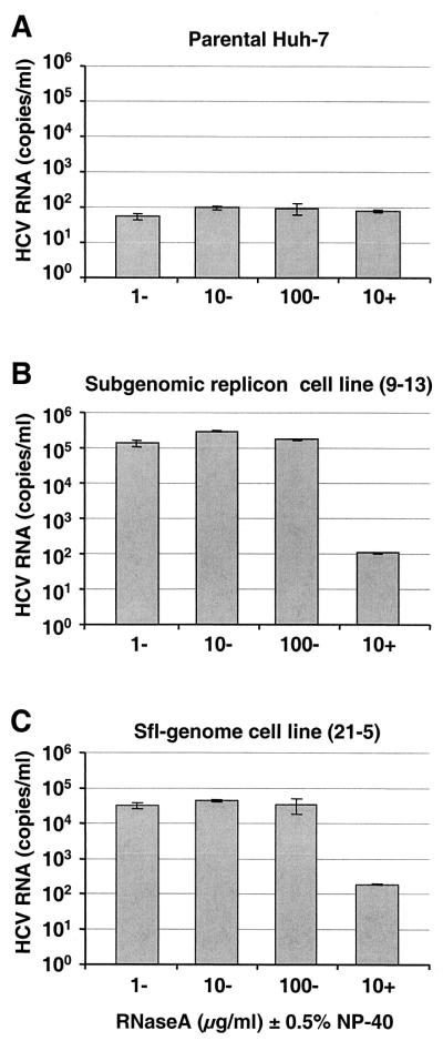
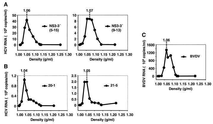
The recently developed subgenomic hepatitis C virus (HCV) replicons were limited by the fact that the sequence encoding the structural proteins was missing. Therefore, important information about a possible influence of these proteins on replication and pathogenesis and about the mechanism of virus formation could not be obtained. Taking advantage of three cell culture-adaptive mutations that enhance RNA replication synergistically, we generated selectable full-length HCV genomes that amplify to high levels in the human hepatoma cell line Huh-7 and can be stably propagated for more than 6 months. The structural proteins are efficiently expressed, with the viral glycoproteins E1 and E2 forming heterodimers which are stable under nondenaturing conditions. No disulfide-linked glycoprotein aggregates were observed, suggesting that the envelope proteins fold productively. Electron microscopy studies indicate that cell lines harboring these full-length HCV RNAs contain lipid droplets. The majority of the core protein was found on the surfaces of these structures, whereas the glycoproteins appear to localize to the endoplasmic reticulum and cis-Golgi compartments. In agreement with this distribution, no endoglycosidase H-resistant forms of these proteins were detectable. In a search for the production of viral particles, we noticed that these cells release substantial amounts of nuclease-resistant HCV RNA-containing structures with a buoyant density of 1.04 to 1.1 g/ml in iodixanol gradients. The same observation was made in transient-replication assays using an authentic highly adapted full-length HCV genome that lacks heterologous sequences. However, the fact that comparable amounts of such RNA-containing structures were found in the supernatant of cells carrying subgenomic replicons demonstrates a nonspecific release independent of the presence of the structural proteins. These results suggest that Huh-7 cells lack host cell factors that are important for virus particle assembly and/or release.


Figures

FIG. 1.
FIG. 1.
Generation of Huh-7 cell lines bearing sfl genomes. (A) Schematic representations of the structures of the HCV genomes and selectable HCV RNAs used in this study. The length of the construct (number of nucleotides) is given in parentheses to the right of each construct. The parental HCV genome Con1 is shown at the top. The proteins of the polyprotein encoded by the viral RNA are shown (open boxes), and cleavage sites between given proteins are indicated by vertical lines. 5′ and 3′ NTRs flanking the polyprotein are indicated (thick lines). The Con1-ET construct is a derivative that carries three cell culture-adaptive mutations (E1202G, T1280I, and K1846T; Lohmann et al., unpublished) indicated by black dots. The selectable HCV RNAs depicted below both contain an internal EMCV IRES (E-I) directing the expression of core to 5B (sfl genome) or NS3 to 5B (replicon-ET). The selectable marker (neo or zeo, respectively) is expressed via the HCV IRES. While replicon-ET carries the same combination of adaptive mutations as Con1-ET, the sfl genome harbors the SfiI fragment (shaded area) derived from the highly adapted subgenomic replicon rep 5.1 (contributing adaptive mutations E1202G, T1280I, and S2197P). Note that the HCV IRES extends into the core coding region and therefore the selectable markers (neo and zeo) are expressed as fusion proteins carrying the initial 12 amino acids of HCV core. (B) Northern blot of cell lines carrying an sfl genome. Total RNA from seven different sfl cell lines (lanes 2 to 8) was prepared, and 2 μg was analyzed by Northern blotting using 32P-labeled riboprobes complementary to a region within the NS5B gene of HCV and β-actin. For a reference, 2 μg of total RNA derived from naive Huh-7 cells (lane 1) or from subgenomic replicon cell line 9-13 (49) (lane 9) as well as serial dilutions of the original in vitro transcripts (subgenomic replicon [lanes 10 to 12] and sfl genome [lanes 13 to 15]) were analyzed in parallel.
FIG. 2.
FIG. 2.
Stability of sfl genomes during continuous cell culture. (A) Six different cell lines were continuously cultured in the presence of G418. At the indicated time points (days [d]), cells were lysed, total RNA was prepared, and 4 μg was analyzed by Northern blotting. For comparison, given amounts of RNA containing subgenomic replicon transcribed in vitro were analyzed in parallel. To allow detection of sequence variants and of smaller HCV RNA pieces, the entire blot was hybridized under low-stringency conditions. (B) Genetic drift of the sfl genome in cell line 21-5. Cells were continuously cultured, and after 57 passages (corresponding to ∼6 months), total RNA was prepared. The coding region of the HCV polyprotein was amplified by long-distance RT-PCR in two overlapping fragments shown as bars below the ORF. For each fragment, two independent clones were sequenced. The boundary of the sequenced clones is indicated by a dashed vertical line. Amino acid substitutions are drawn as vertical lines at their respective positions within the HCV polyprotein. The hypervariable region 1 (HVR1) present at the N terminus of E2 is depicted as a shaded area. Six mutations were conserved between both sequenced clones, and they are indicated (stars).
FIG. 3.
FIG. 3.
Analysis of structural protein expression in cell lines carrying sfl genomes. (A) Detection of HCV core protein by Western blotting. Lysates, each corresponding to 2 × 10⁵ cells of the given cell lines, were loaded onto a gel, separated by electrophoreses, blotted, and probed with an HCV core-specific monoclonal antibody. (B) Pulse-chase and endoglycosidase H analysis of HCV glycoprotein complexes. Cells were incubated with [35S]methionine/cysteine-containing medium for 1 h, and after the cells were washed extensively, nonradioactive medium was added for the times specified above the lanes. Glycoprotein complexes were isolated by immunoprecipitations from cell lysates under nondenaturing conditions and were either mock treated (− EndoH) or deglycosylated by endoglycosidase H digestion (+ EndoH). After SDS-PAGE, proteins were detected by autoradiography. The deglycosylated forms of the proteins are labeled with asterisks. (C) Analysis of HCV glycoprotein complexes under reducing and nonreducing conditions. Immunoprecipitations were performed in the presence of 20 mM iodoacetamide in the lysis and wash buffer. Prior to loading, half of each sample was boiled in denaturing buffer with or without 2-ME as indicated. Proteins were detected as described above.
FIG. 4.
FIG. 4.
Morphology of cell lines with an sfl genome and localization of viral proteins. (A) Ultrathin section obtained from cell line 21-5 after glutaraldehyde-osmium fixation and ERL embedding. Magnification, ×28,500. The cells exhibit numerous lipid vesicles (V) of different sizes surrounded to different degrees by electron-dense rims resembling a thickened membrane. (B) Ultrathin section of parental Huh-7 cells (fixation, embedding, and magnification as for panel A). Note the absence of the electron-dense rim around the border of the vesicles. (C to E) Localization of HCV E2 (C) and core (D) proteins and the Golgi-resident protein Golgin 97 (E) in cell line 21-5 by indirect immunofluorescence. Representative sections are shown at a magnification of ×304. Bar, 50 μm. Nuclei were counterstained using bisbenzimide (Hoechst). Note the granular staining pattern of the core protein that to some extent resembles the distribution of the Golgi marker Golgin 97. (I and J) Specificity control for the anti-core and anti-E2 sera. Cells harboring a subgenomic replicon were fixed and stained as cells with the sfl genome. Magnification, ×285. (F to H) Localization of E2, core, and Golgin 97 in cell line 21-5 treated with 20 nM brefeldin A for 3 h. Detection of Golgin 97 is abrogated (H), while the distribution of E2 and core remains essentially unchanged (F and G, respectively). Magnification, ×304.
FIG. 5.
FIG. 5.
Subcellular localization of HCV proteins by immuno-EM. (A and B) Localization of HCV core (6-nm-diameter gold particles) and E2 (10-nm-diameter gold particles) in cell line 21-5. (A) The majority of the core protein is found on the surfaces of lipid vesicles (V), and only minor staining is detected at the ER. No 10-nm-diameter gold specific for E2 can be seen in the area around the vesicles. Note that no osmium postfixation was used, and therefore, the lipid vesicles appear empty, due to removal of the content by alcohol dehydration and embedding medium. (B) The 10-nm-diameter gold marker specific for E2 associated with elongated, cisterna-like structures in the cytoplasm presumably belonging to the ER and early compartments of the Golgi complex. Note the absence of 6-nm-diameter gold marker specific for core. (C) Specificity control of the immuno-gold labeling. Cell line 21-5 was labeled with primary antibodies directed against HIV and measles virus proteins, which share the isotypes of the antibodies used in panels A and B. Subsequently, the same gold-labeled secondary antibodies were used. Magnification, ×92,150.
FIG. 6.
FIG. 6.
Detection of nuclease-resistant HCV-RNAs in supernatants of various cell lines. Supernatants from parental Huh-7 cells (A), cell line 9-13 harboring a subgenomic replicon (B), and cell line 21-5 with an sfl genome (C) were harvested 6 days postseeding, cleared by low-speed centrifugation, filtered through 0.45-μm-pore-size filters, and pelleted by high-speed ultracentrifugation. Pellets were resuspended in PBS supplemented with carrier RNA, and aliquots were incubated for 4 h at room temperature with RNase A at the concentrations given below the bars. Some aliquots were also supplemented with 0.5% NP-40 (indicated by + [−, no NP-40 added]). HCV RNA was measured by quantitative RT-PCR performed in duplicate. Mean values of duplicates representing the number of HCV RNA molecules per milliliter of supernatant are given, and the error bars indicate the standard errors of the means.
FIG. 7.
FIG. 7.
Analysis of released HCV RNA by density gradient centrifugation. (A and B) Cell culture supernatants from two different cell lines carrying subgenomic replicons (5-15 and 9-13) (A) and two cell lines carrying sfl genomes (20-1 and 21-5) (B) were harvested 3 days postseeding and processed as described in Materials and Methods. RNA was prepared and subjected to quantitative RT-PCR. (C) For a control, supernatant from R.D.420 cells productively infected with BVDV (strain NADL) was analyzed in parallel. Copies of HCV and BVDV RNA detected per milliliter of gradient fraction (mean value of duplicates) are plotted against the density. The density of the peak fraction is given at the top of each graph.
FIG. 8.
FIG. 8.
Transient replication of a subgenomic replicon and full-length HCV genomes. Huh-7 cells were transfected with the authentic HCV Con1 genome (Con1), a cell culture-adapted full-length RNA (Con1-ET), or a selectable subgenomic replicon (replicon-ET), and cells were harvested at the indicated time points. (A and B) Transient replication was monitored by Northern blotting (A), as described in the legend to Fig. 1B, and NS5B-specific Western blotting (B). (C) At 96 h posttransfection, supernatants from three 10-cm-diameter dishes were harvested, cleared of cellular debris, and subjected to density gradient centrifugation as described in Materials and Methods. HCV RNA in each fraction was measured by quantitative RT-PCR. The average value of duplicate experiments is plotted against the density of the corresponding fraction.

References

    1. Barba, G., F. Harper, T. Harada, M. Kohara, S. Goulinet, Y. Matsuura, G. Eder, Z. Schaff, M. J. Chapman, T. Miyamura, and C. Brechot. 1997. Hepatitis C virus core protein shows a cytoplasmic localization and associates to cellular lipid storage droplets. Proc. Natl. Acad. Sci. USA 94:1200-1205. - PMC - PubMed
    1. Barbaro, G., G. Di Lorenzo, A. Asti, M. Ribersani, G. Belloni, B. Grisorio, G. Filice, and G. Barbarini. 1999. Hepatocellular mitochondrial alterations in patients with chronic hepatitis C: ultrastructural and biochemical findings. Am. J. Gastroenterol. 94:2198-2205. - PubMed
    1. Bartenschlager, R. 1999. The NS3/4A proteinase of the hepatitis C virus: unravelling structure and function of an unusual enzyme and a prime target for antiviral therapy. J. Viral Hepatitis 10:1-2. - PubMed
    1. Bartenschlager, R., L. L. Ahlborn, J. Mous, and H. Jacobsen. 1993. Nonstructural protein 3 of the hepatitis C virus encodes a serine-type proteinase required for cleavage at the NS3/4 and NS4/5 junctions. J. Virol. 67:3835-3844. - PMC - PubMed
    1. Bartenschlager, R., and V. Lohmann. 2000. Replication of hepatitis C virus. J. Gen. Virol. 81:1631-1648. - PubMed

Publication types