. 2002 Apr;76(8):4044-55.
doi: 10.1128/jvi.76.8.4044-4055.2002.

Structural requirements for the assembly of Norwalk virus-like particles

Affiliations

Structural requirements for the assembly of Norwalk virus-like particles

Andrea Bertolotti-Ciarlet et al. J Virol. 2002 Apr.

Abstract

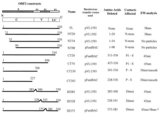
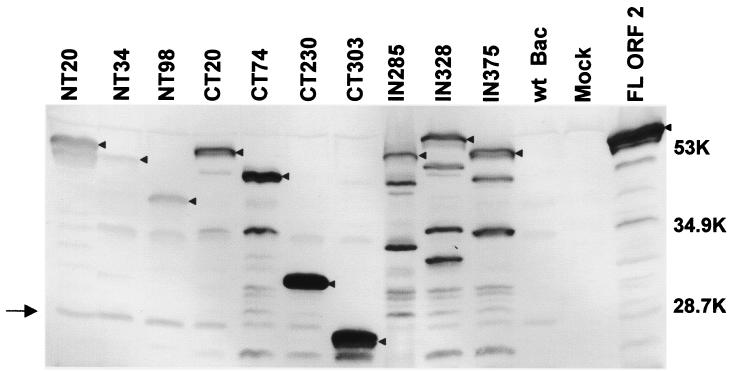
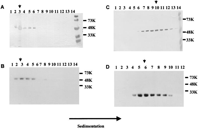
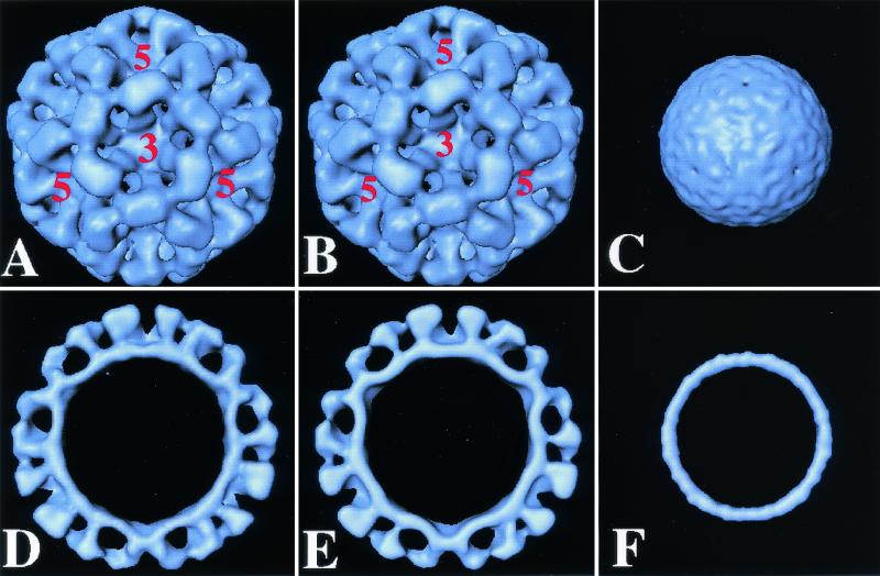
Norwalk virus (NV) is the prototype strain of a group of human caliciviruses responsible for epidemic outbreaks of acute gastroenteritis. While these viruses do not grow in tissue culture cells or animal models, expression of the capsid protein in insect cells results in the self-assembly of recombinant NV virus-like particles (rNV VLPs) that are morphologically and antigenically similar to native NV. The X-ray structure of the rNV VLPs has revealed that the capsid protein folds into two principal domains: a shell (S) domain and a protruding (P) domain (B. V. V. Prasad, M. E. Hardy, T. Dokland, J. Bella, M. G. Rossmann, and M. K. Estes, Science 286:287-290, 1999). To investigate the structural requirements for the assembly of rNV VLPs, we performed mutational analyses of the capsid protein. We examined the ability of 10 deletion mutants of the capsid protein to assemble into VLPs in insect cell cultures. Deletion of the N-terminal 20 residues, suggested by the X-ray structure to be involved in a switching mechanism during assembly, did not affect the ability of the mutant capsid protein to self-assemble into 38-nm VLPs with a T=3 icosahedral symmetry. Further deletions in the N-terminal region affected particle assembly. Deletions in the C-terminal regions of the P domain, involved in the interactions between the P and S domains, did not block the assembly process, but they affected the size and stability of the particles. Mutants carrying three internal deletion mutations in the P domain, involved in maintaining dimeric interactions, produced significantly larger 45-nm particles, albeit in low yields. The complete removal of the protruding domain resulted in the formation of smooth particles with a diameter that is slightly smaller than the 30-nm diameter expected from the rNV structure. These studies indicate that the shell domain of the NV capsid protein contains everything required to initiate the assembly of the capsid, whereas the entire protruding domain contributes to the increased stability of the capsid by adding intermolecular contacts between the dimeric subunits and may control the size of the capsid.


Figures

FIG. 1.
FIG. 1.
Schematic representation of the constructs used in this study to produce NV capsid mutants using the baculovirus expression system. The left column shows a schematic representation of the NV capsid protein and each mutant. The conserved (C), variable (V), and less-conserved (LC) regions based on sequence identity among Norwalk-like viruses are shown within the schematic of the full-length (FL) capsid protein. Above this schematic representation, the amino acid sequence that relates to each domain of the capsid structure is indicated. The figure also shows the transfer vector used for expression, which amino acids were deleted, and what contacts were predicted to be affected. The right column shows the size of particles observed by electron microscopy when the particles were purified over iodixanol or sucrose gradients. ∗ indicates that the 38-nm particles were observed only when particles were purified over iodixanol gradients. Mutant NT34 was incorrectly called NT35 when first described (47).
FIG. 2.
FIG. 2.
Expression of NV mutant capsid proteins. Infected cell lysates (3 × 10⁶ cells) were harvested at 36 hpi in 300 μl of 5× SDS-PAGE sample buffer (3 parts disruption buffer [10% SDS, 50% 2-mercaptoethanol, 5 M urea, 1:1:1]; 2 parts F/2 [1:1; stacking gel buffer {0.5 M Tris-HCl, 0.46% TEMED, pH 6.6 to 6.8}), 80% glycerol; 0.02 g of phenol red]) and analyzed by SDS-12% PAGE and Western blotting. In order to compare the relative levels of protein expression of the mutant proteins, the same volume of lysate was loaded for each mutant. Western blotting was performed using a rabbit hyperimmune serum to rNV VLPs (27). At the dilution used, this antiserum also recognizes one nonspecific band in the wild-type-baculovirus-infected cell lysates at around 20 kDa (→). Although protease inhibitors were added to the infected cells, some degradation products were observed for all the constructs. Arrowheads indicate the bands that correspond to the uncleaved mutant or full-length capsid proteins. The levels of expression of each mutant were quantitated by ELISA (see text). Sizes are shown in kilodaltons.
FIG. 3.
FIG. 3.
Analysis of the presence of capsid protein in fractions of iodixanol and sucrose gradients. The supernatant of infected cells (3 × 10⁶ cells/ml in 200 ml) was sedimented and separated through sucrose or iodixanol gradients. The figure shows Western blotting analysis of fractions from iodixanol (A and B) and sucrose (C and D) gradients. The fractions shown to contain capsid protein were pooled, and the sucrose or iodixanol was removed by dilution in phosphate buffer and sedimentation. (A) Full-length NV capsid, (B) NT20, (C) ID375, and (D) CT230. Sizes are shown in kilodaltons. The arrowhead shows the peak fraction in each gradient.
FIG. 4.
FIG. 4.
Electron microscopic analysis of NV capsid mutants. For each mutant, the top panel shows the region mutated (red) on a rope representation (white) of the full-length NV capsid protein crystal structure (38), and the lower panel shows an electron micrograph of the different mutant particles purified from undiluted supernatant material from the Sf9 cell cultures. The electron micrographs show a representative area of grids prepared with gradient fractions. Ammonium molybdate (1%) was used for staining. (A) NT20 forms VLPs that resemble full-length capsid protein VLPs. (B and C) CT20 and CT74 form VLPs that are 45 nm in diameter. (D and E) CT230 and CT303 form VLPs that lack the characteristic arches formed by the protruding domains and resemble smooth particles. (F and G) ID285 and ID328 form VLPs that are 45 nm in diameter. (H) ID375 forms VLPs that are 45 nm in diameter, as well as VLPs that are morphologically similar to full-length capsid protein VLPs when purified over iodixanol gradients. Bar, 50 nm.
FIG. 5.
FIG. 5.
Comparison of the structure of full-length and mutant NT20 and CT303 rNV VLPs. Surface representation of the cryoelectron microscopic reconstruction of full-length rNV VLPs (A) at 22 Å and its cross-section (D), the NT20 mutant structure (B) and its cross-section (E), and the CT303 mutant structure (C) and its cross-section (F). The five- and threefold axes are indicated.

References

    1. Crowther, R. A. 1971. Procedures for three-dimensional reconstruction of spherical viruses by Fourier synthesis from electron micrographs. Phil. Trans. R. Scot. Lond. B Biol. Sci. 261:221-230. - PubMed
    1. Crowther, R. A., D. J. DeRosier, and A. Klug. 1970. The reconstruction of a three-dimensional structure from projections and its application to electron microscopy. Proc. R. Soc. Lond. 317:319. - PubMed
    1. Delgadillo, R. A., R. M. Smets, Q. R. Yang, D. Vanden Berghe, and A. Neetens. 1988. The use of iodinated density gradient medium for the isolation of rod outer segments. Experientia 44:702-704. - PubMed
    1. Dong, X. F., P. Natarajan, M. Tihova, J. E. Johnson, and A. Schneemann. 1998. Particle polymorphism caused by deletion of a peptide molecular switch in a quasiequivalent icosahedral virus. J. Virol 72:6024-6033. - PMC - PubMed
    1. Dubochet, J., M. Adrian, J. J. Chang, J. C. Homo, J. Lepault, A. W. McDowall, and P. Schultz. 1988. Cryo-electron microscopy of vitrified specimens. Q. Rev. Biophys. 21:129-228. - PubMed

Publication types

LinkOut - more resources