. 2002 Apr;76(8):4080-6.
doi: 10.1128/jvi.76.8.4080-4086.2002.

Preferential localization of the Epstein-Barr virus (EBV) oncoprotein LMP-1 to nuclei in human T cells: implications for its role in the development of EBV genome-positive T-cell lymphomas

Affiliations

Preferential localization of the Epstein-Barr virus (EBV) oncoprotein LMP-1 to nuclei in human T cells: implications for its role in the development of EBV genome-positive T-cell lymphomas

Jingwu Xu et al. J Virol. 2002 Apr.

Abstract

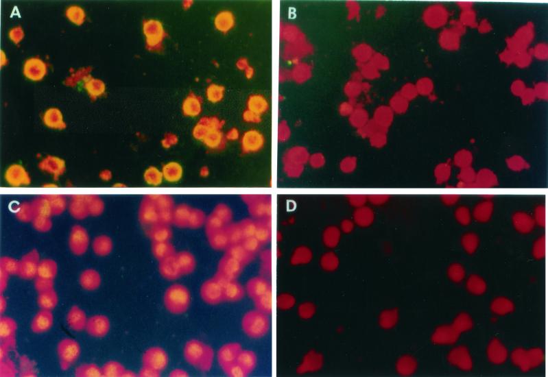
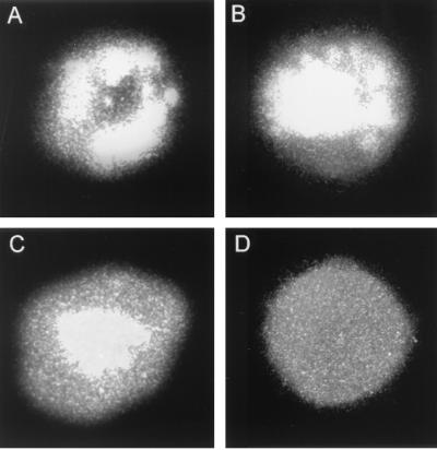
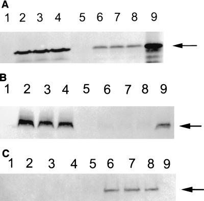
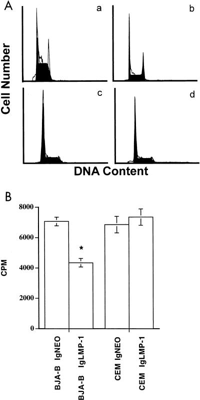
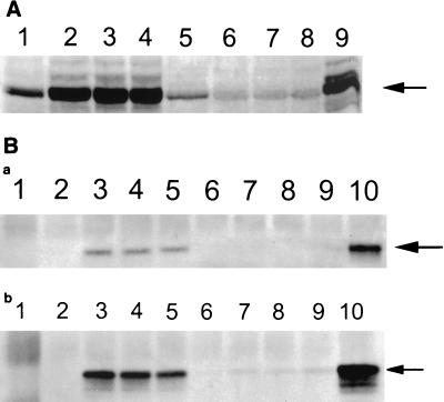
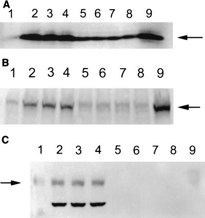
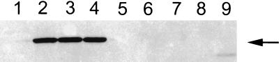
The Epstein-Barr virus (EBV)-encoded latent membrane protein-1 (LMP-1) is thought to play a role in the EBV-induced B-cell transformation and immortalization. EBV has also been implicated in certain human T-cell lymphomas; however, the phenotypic effects of the expression of this oncoprotein in T cells are not known. To learn whether LMP-1 also induces phenotypic changes in T cells, we stably expressed it in human cell lines of T and B lineages and 25 LMP-1-expressing T-cell clones and 7 B-cell clones were examined. Our results show for the first time that, in sharp contrast to B cells, LMP-1 preferentially localizes to nuclei in T cells and does not induce the phenotypic changes in these cells that it induces in B cells, does not associate with TRAF proteins, and does not arrest the cell cycle in the G2/M phase. A computer-assisted analysis revealed that LMP-1 lacks the canonical nuclear localization signal. Our results suggest that this oncoprotein may not play the same role in the lymphomagenesis of T cells as it does in B cells.


Figures

FIG. 1.
FIG. 1.
Expression of LMP-1 in CEM and BJA-B cells. LMP-1 was detected by indirect immunofluorescence in permeabilized cells with S12 antibody and fluorescein isothiocyanate-conjugated goat anti-mouse IgG. The upper panels show LMP-expressing BJA-B cells stained with S12 (A) or control (B) antibody. The lower panels show LMP-expressing CEM cells stained with S12 (C) or control (negative) (D) antibody. Note the localization of immunofluorescence in the nucleus in CEM cells (C) and at the membranes in BJA-B cells (A).
FIG. 2.
FIG. 2.
Different patterns of LMP-1 localization in Molt4 cells. LMP-expressing Molt4 cells were stained with S12 as described in the legend to Fig. 1. Shown here are three different patterns of LMP-1 localization in these cells: on the membrane (A), both on the membrane and in the nucleus (B), and in the nucleus (C). Panel D represents an LMP-expressing cell stained with a control (negative) antibody.
FIG. 3.
FIG. 3.
Detection of LMP-1 by Western blotting in CEM and BJA-B cells. (A) Whole-cell lysates (40 μg) from LMP-expressing and control vector-transfected cells were resolved by SDS-10% PAGE. After transfer onto nylon membranes, LMP-1 was detected by anti-LMP monoclonal antibody S12 followed by detection with alkaline phosphatase-conjugated anti-mouse antibodies 5-bromo-4-chloro-3-indolylphosphate (BCIP) and nitroblue tetrazolium. Lanes: 1, BJA-B transfected with the control vector; 2 to 4, three different LMP-expressing BJA-B clones; 5, CEM transfected with the control vector; 6 to 8, three different LMP-expressing CEM clones; 9, an EBV-transformed B-cell line (LCL). The LMP-expressing BJA-B and CEM cells were fractionated into cytoplasmic and nuclear lysates, and each fraction was analyzed for LMP-1 expression by Western blotting. Panels B and C show LMP-1 expression in cytoplasmic and nuclear fractions, respectively. The lanes represent the clones indicated for panel A. The arrows point to LMP-1 bands.
FIG. 4.
FIG. 4.
Cell cycle profiles of LMP-expressing cells. After staining with PI, the cell cycle profile was analyzed from the DNA content by using a flow cytometer and Cellfit software. (A) The figure shows the relative number of cells in various phases of the cell cycle for control vector-transfected BJA-B (a), LMP-expressing BJA-B (b), control vector-transfected CEM (c), and LMP-expressing CEM (d) cells. Note the accumulation of cells in the G2/M phase in LMP-expressing BJA-B cells (b). (B) Proliferation of pIgLMP-1 and control vector (IgNEO)-transfected cells. Cells (105 per well) were cultured in triplicate for 24 h and pulsed with [3H]TdR for 16 h to assess proliferation. The asterisk indicates significant difference (P < 0.01).
FIG. 5.
FIG. 5.
(A) LMP-1 induced the TRAF1 protein (arrow) in BJA-B cells but not in CEM cells. Whole-cell lysates (40 μg) from LMP-expressing and control vector-transfected cells were resolved by SDS-10% PAGE. After transfer onto nylon membranes, TRAF1 was incubated with anti-TRAF1 antibody followed by detection with alkaline phosphatase-conjugated anti-rabbit antibody. The lanes represent different clones (the same as lanes 1 to 9 of Fig. 3). (B) LMP-1 (arrows) interacted with TRAF1 in BJA-B cells but not in CEM cells. TRAF1 was immunoprecipitated from cytoplasmic fractions (a) and whole-cell lysates (b), resolved by SDS-PAGE, and electroblotted onto nylon membranes. The membranes were probed for LMP-1 with LMP-specific monoclonal antibodies (CS1-4) and alkaline phosphatase-conjugated secondary antibodies. Lane 1 represents immunoprecipitation with normal rabbit serum. Lanes 2 to 10 in panel B correspond to lanes 1 to 9 in panel A, respectively.
FIG. 6.
FIG. 6.
Expression of bcl-2 (A), LFA-1 (B), and ICAM-1 (C). LMP expression induces bcl-2, LFA-1, and ICAM-1 in BJA-B cells but not in CEM cells. The expression of these proteins in different clones of LMP-expressing cells was determined by Western blotting performed on 40 μg of whole-cell lysates. The lanes represent the cell clones indicated in the legend to Fig. 3. Arrows from top to bottom indicate bcl-2, LFA-1, and ICAM-1, respectively.
FIG. 7.
FIG. 7.
LMP-1 expression induces a tyrosine-phosphorylated protein in B, but not in T, cells (arrow). Far Western blots were prepared from whole-cell lysates by using immunoprecipitated LMP-1. The blots were probed with anti-phosphotyrosine and alkaline phosphatase-conjugated anti-mouse IgG as described in the legend to Fig. 3. The lanes indicate the cell clones described in the legend to Fig. 3.

References

    1. Ahmad, A., A. Ladha, E. A. Cohen, and J. Menezes. 1993. Stable expression of the transfected HIV-1 env gene in a human B cell line: characterization of gp120-expressing clones and immunobiological studies. Virology 192:447-457. - PubMed
    1. Arvanitakis, L., N. Yaseen, and S. Sharma. 1995. Latent membrane protein-1 induces cyclin D2 expression, pRb hyperphosphorylation, and loss of TGF-beta 1-mediated growth inhibition in EBV-positive B cells. J. Immunol. 155:1047-1056. - PubMed
    1. Babcock, G. J., L. L. Decker, M. Volk, and D. A. Thorley-Lawson. 1998. EBV persistence in memory B cells in vivo. Immunity 9:395-404. - PubMed
    1. Birkenbach, M., D. Liebowitz, F. Wang, J. Sample, and E. Kieff. 1989. Epstein-Barr virus latent infection membrane protein increases vimentin expression in human B-cell lines. J. Virol. 63:4079-4084. - PMC - PubMed
    1. Bonagura, V. R., B. Z. Katz, B. C. Edwards, D. J. Valaces, P. Nisen, E. Gloster, R. Mir, and P. Lanzkowsky. 1990. Severe chronic EBV infection associated with specific EBV immunodeficiency and an EBNA+ T-cell lymphoma containing linear, EBV DNA. Clin. Immunol. Immunopathol. 57:32-44. - PubMed

Publication types

Substances