. 2002 Apr;22(8):2427-40.
doi: 10.1128/MCB.22.8.2427-2440.2002.

SRC catalytic but not scaffolding function is needed for integrin-regulated tyrosine phosphorylation, cell migration, and cell spreading

Affiliations

SRC catalytic but not scaffolding function is needed for integrin-regulated tyrosine phosphorylation, cell migration, and cell spreading

Leslie A Cary et al. Mol Cell Biol. 2002 Apr.

Abstract

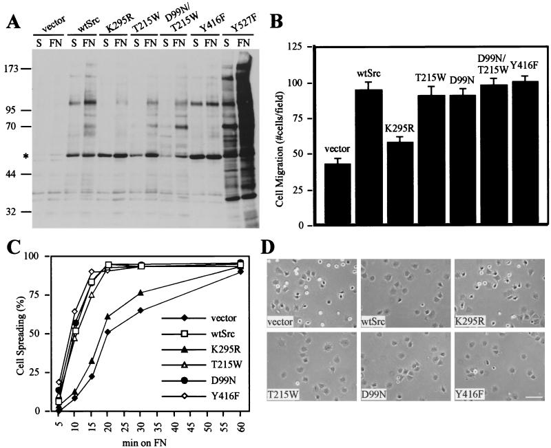
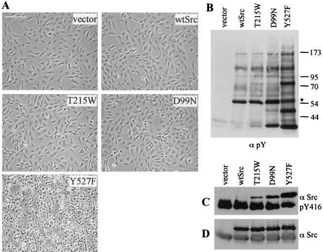
Src family kinases (SFKs) are crucial for signaling through a variety of cell surface receptors, including integrins. There is evidence that integrin activation induces focal adhesion kinase (FAK) autophosphorylation at Y397 and that Src binds to and is activated by FAK to carry out subsequent phosphorylation events. However, it has also been suggested that Src functions as a scaffolding molecule through its SH2 and SH3 domains and that its kinase activity is not necessary. To examine the role of SFKs in integrin signaling, we have expressed various Src molecules in fibroblasts lacking other SFKs. In cells plated on fibronectin, FAK could indeed autophosphorylate at Y397 independently of Src but with lower efficiency than when Src was present. This step was promoted by kinase-inactive Src, but Src kinase activity was required for full rescue. Src kinase activity was also required for phosphorylation of additional sites on FAK and for other integrin-directed functions, including cell migration and spreading on fibronectin. In contrast, Src mutations in the SH2 or SH3 domain greatly reduced binding to FAK, Cas, and paxillin but had little effect on tyrosine phosphorylation or biological assays. Furthermore, our indirect evidence indicates that Src kinase activity does not need to be regulated to promote cell migration and FAK phosphorylation. Although Src clearly plays important roles in integrin signaling, it was not concentrated in focal adhesions. These results indicate that the primary role of Src in integrin signaling is as a kinase. Indirect models for Src function are proposed.


Figures

FIG. 1.
FIG. 1.
Integrin-induced phosphorylation, migration and spreading require catalytic but not scaffolding functions of Src. SYF cells reconstituted with vector, wtSrc, or the indicated Src mutants (see Results for descriptions) were harvested with trypsin, washed, and resuspended in serum-free medium. (A) Cells were lysed with RIPA buffer in suspension (S) or after plating on fibronectin-coated dishes for 30 min (FN). Total cell lysates were Western blotted with the anti-phosphotyrosine MAb 4G10. The position of molecular mass markers (in kDa) is shown on the left. An asterisk indicates the position of Src. (B) Cell migration towards 10 μg of fibronectin/ml was measured using a chemotaxis chamber. Results shown were compiled from three individual experiments for each cell line. Error bars represent standard errors of the mean. No cells migrated when DMEM without FN was added to the lower wells (not shown). (C and D) Cells were plated on FN-coated dishes at 37°C, and time-lapse phase-contrast images were taken at the indicated times. This experiment was repeated at least three times with similar results. (C) The percentages of cells that had spread at each time point were scored as indicated. (D) Representative images taken 20 min after cell plating. Scale bar, ∼100 μm.
FIG. 2.
FIG. 2.
Src kinase activity is needed for integrin-dependent FAK phosphorylation. SYF cells were infected using a retroviral system to stably express wtSrc or KD-Src mutants (K295R or D386A) or were infected with virus containing empty vector. Cells were harvested with trypsin, washed, and lysed with Triton buffer in suspension (0 min) or after plating for the indicated times (10, 20, or 30 min) on FN-coated dishes. FAK tyrosine phosphorylation was examined by immunoprecipitating with anti-FAK C20, followed by Western blotting with either anti-FAK C20 to demonstrate equal amounts of FAK in each immunoprecipitate (A), anti-phosphotyrosine 4G10 (B), or the site-specific phosphoantibodies anti-FAK-pY576 (C) or anti-FAK-pY397 (D). Src expression levels were determined by Western blotting cell lysates with anti-Src LA074 (E).
FIG. 3.
FIG. 3.
Regulated Src kinase activity is not required for FAK phosphorylation upon integrin activation. SYF cells expressing the indicated Src molecules (or reconstituted with empty vector) were harvested with trypsin, washed, and lysed in suspension (0 min) or after plating on FN-coated dishes for 10 or 30 min, as indicated. (A and B) Src was immunoprecipitated from RIPA buffer lysates with anti-Src LA074, followed by Western blotting with anti-Src-pY416 (A) or anti-Src SRC2 (B). (C and D) FAK was immunoprecipitated from Triton buffer lysates with anti-FAK C20 followed by Western blotting with anti-phosphotyrosine 4G10 (C) or anti-FAK C20 (D).
FIG. 4.
FIG. 4.
Src is not localized to focal adhesions, while localization of other focal adhesion proteins is independent of SFK expression. SYF cells reconstituted with vector (B and D), wtSrc (A, C, E, and G), or Y416F Src mutant (F and H) were harvested with trypsin, washed, and plated on FN-coated coverslips in 0.5% FBS for 1 h. Cells were washed, fixed, permeabilized and stained with MAb anti-FAK (A and B) or anti-Src LA074 (C and D), followed by FITC-conjugated anti-mouse secondary antibody. Some cells were costained with MAb anti-paxillin (E and F) and anti-Src pY416 (G and H), followed by Texas Red and FITC-conjugated secondary antibodies to detect paxillin and active Src, respectively. The faint nuclear staining seen with anti-Src pY416 (G and H) is due to nonspecific bleed-through from DAPI staining (not shown). Scale bar, ∼50 μm.
FIG. 5.
FIG. 5.
Activity of Src SH2 and SH3 domain mutants. SYF cells stably expressing wtSrc, an SH2 domain mutant (T215W) or an SH3 domain mutant (D99N), or an activated mutant (Y527F) were established. (A) Phase-contrast images of near-confluent cell cultures at ×100 magnification (scale bar, ∼200 μm). (B) Lysates using Triton buffer were generated from similar cultures, and total proteins were Western blotted with anti-phosphotyrosine MAb 4G10. The positions of molecular mass markers (in kDa) are shown on the right. An asterisk indicates the position of Src. To detect the activation level of each form of Src, anti-Src immunoprecipitates (using MAb LA074) from growing cells were Western blotted with anti-Src-pY416 (C) or anti-Src LA074 (D).
FIG. 6.
FIG. 6.
Src SH2 and SH3 domain mutations reduce binding to FAK, paxillin, and Cas. (A and B) NIH3T3 cells were lysed on the dish (Att) or were harvested with trypsin and lysed in suspension (Sus). These lysates were used for in vitro binding experiments with GST-Src fusion proteins. (A) Levels of FAK and paxillin input in RIPA buffer cell lysates (not shown) or immunoprecipitates (left) were equal. Attachment-induced phosphorylation of immunoprecipitated FAK and paxillin was detected with MAb 4G10 (middle). Attachment-induced binding of FAK and paxillin to GST-SrcSH2 was significantly reduced by the T215W mutation (right). (B) Binding of FAK from Triton buffer lysates to GST-SrcSH3 was not affected by FAK phosphotyrosine levels (left). Binding of FAK (middle) and Cas (right) (from attached cells) to GST-SrcSH3 was significantly reduced by the D99N mutation. The input level of Cas shown (right) is 5% of that used for the binding experiment. (C) Approximately equal amounts of each GST fusion protein were prepared as determined by Coomassie staining, although amounts used for binding assays were adjusted slightly to account for differences. (D through G) SYF cells reconstituted with vector or the indicated Src molecules were harvested with trypsin, washed, and lysed with NP-40 buffer in suspension (Sus) or after replating on FN-coated dishes for 30 min (FN). Whole cell lysates were Western blotted with anti-FAK C20 (D) or anti-Src LA074 (E). Alternatively, Src was immunoprecipitated with LA074 followed by Western blotting with anti-FAK C20 to detect associated FAK (F) or anti-Src LA074 (G). WB, Western blotting; IP, immunoprecipitation.
FIG. 7.
FIG. 7.
Src-mediated FAK, Cas, and paxillin phosphorylation does not require stable association. SYF cells were reconstituted with vector, wtSrc, or the indicated Src mutants (T215W, D99N, D99N/T215W, or Y527F). Cells were harvested with trypsin, washed, and lysed in suspension (S) or after replating on FN-coated dishes (FN) for 30 (A through D) or 15 (E through H) min. (A through D) FAK was immunoprecipitated from Triton buffer lysates with anti-FAK C20, followed by Western blotting with anti-phosphotyrosine 4G10 (A) or with anti-FAK C20 (B). Some immunoprecipitates were also Western blotted with the site-specific phosphorylation antibody anti-FAK-pY397 (C) or anti-FAK-pY576 (D). Cas (E and F) or paxillin (G and H) immunoprecipitates from RIPA buffer lysates were Western blotted with 4G10 (E and G), anti-Cas (F), or anti-paxillin (H).
FIG. 8.
FIG. 8.
Src scaffolding functions can occur when KD-Src is overexpressed. (A) SYF cells expressing low or high levels of wtSrc or KD-Src (K295R mutant) were established as described in Materials and Methods. Src expression levels were compared with that of endogenous Src in fibroblasts derived from a src+/+fyn−/− yes−/− (src+/+) mouse embryo. Src expression levels were determined by Western blotting cell lysates (using Triton buffer) with anti-Src LA074 (left) or SRC2 (right). (B through E) Cells expressing low or high levels of wtSrc or KD-Src were harvested with trypsin, washed, and lysed with Triton buffer in suspension (0 min) or after plating on FN-coated dishes for 30 min. (B and C) FAK was immunoprecipitated with anti-KC, followed by Western blotting with anti-phosphotyrosine 4G10 (B) or anti-KC (C). Cas was immunoprecipitated with anti-Cas C20, followed by Western blotting with 4G10 (D) or anti-Cas C20 (E). (F) The cells described above were harvested with Triton lysis buffer in suspension (S) or after plating on FN for 20 min (FN). FAK immunoprecipitates (using anti-FAK C20) were used for in vitro kinase assays using [γ-32P]ATP and poly(Glu, Tyr) as a substrate, and kinase activity was normalized against FAK levels (not shown). (G and H) SYF cells lacking or expressing wtSrc were treated with 0 μM (−) or 50 μM (+) sodium vanadate for 5 h to inhibit tyrosine phosphatases. Cells were lysed (with Triton buffer) attached (A) or were harvested with trypsin, washed, and lysed in suspension (S) or after plating on FN for 40 min (FN). (G) FAK immunoprecipitates (using anti-KC) were Western blotted with 4G10. Equal amounts of FAK were detected in each immunoprecipitate (not shown). (H) Whole cell lysates from wtSrc-expressing cells (lysed attached) were Western blotted with 4G10 to demonstrate vanadate-induced phosphotyrosine of many proteins. The positions of molecular mass markers (in kDa) are shown on the right.
FIG. 9.
FIG. 9.
Models of Src-regulated events in response to integrin activation. Shown here are (A) a conventional model based on previous data describing the roles of Src and FAK in integrin-mediated events and (B and C) two new potential models based on the data presented here. Filled circles (•) represent phosphotyrosine residues, and dashed lines indicate binding interactions. See the text for further explanation of these models. For simplicity, we are showing only one of Src's presumed substrates, Cas, although these models could apply to other proteins as well. (A) Previous work predicted that upon integrin activation (e.g., by FN stimulation), FAK would autophosphorylate at Y397 (1), to allow (2) recruitment of Src through binding its SH2 and SH3 domains and subsequent activation of Src catalytic activity. Src would then phosphorylate additional sites on FAK (3) and associated proteins (4), to promote events such as cell migration or spreading. Because our data indicate that neither regulation of Src catalytic activity nor SH2/SH3-mediated scaffolding functions are required, this model may be correct, except that only weak and transient binding interactions are needed. In an alternative model (B), FAK pY397 is required not for binding Src but for regulating an intermediate protein, X (e.g., a phosphatase), which allows substrates phosphorylated by basally active Src to accumulate locally. A third possibility (C) is that basal Src catalytic activity is sufficient for phosphorylation of protein Y; either Y allows FAK to act as the important kinase (e.g., Y is a phosphatase that, when inhibited by Src, allows FAK to function) or perhaps Y is itself the kinase that carries out these reactions.

References

    1. Angers-Loustau, A., J. F. Cote, and M. L. Tremblay. 1999. Roles of protein tyrosine phosphatases in cell migration and adhesion. Biochem. Cell Biol. 77:493-505. - PubMed
    1. Avraham, H., S. Y. Park, K. Schinkmann, and S. Avraham. 2000. RAFTK/Pyk2-mediated cellular signalling. Cell. Signal. 12:123-133. - PubMed
    1. Boerner, R. J., D. B. Kassel, S. C. Barker, B. Ellis, P. DeLacy, and W. B. Knight. 1996. Correlation of the phosphorylation states of pp60c-src with tyrosine kinase activity: the intramolecular pY530-SH2 complex retains significant activity if Y419 is phosphorylated. Biochemistry 35:9519-9525. - PubMed
    1. Brown, M. T., J. Andrade, H. Radhakrishna, J. G. Donaldson, J. A. Cooper, and P. A. Randazzo. 1998. ASAP1, a phospholipid-dependent Arf GTPase-activating protein that associates with and is phosphorylated by Src. Mol. Cell. Biol. 18:7038-7051. - PMC - PubMed
    1. Brown, M. T., and J. A. Cooper. 1996. Regulation, substrates and functions of src. Biochim. Biophys. Acta 1287:121-149. - PubMed

Publication types

MeSH terms