. 2002 Apr;22(8):2586-97.
doi: 10.1128/MCB.22.8.2586-2597.2002.

Cripto-1 activates nodal- and ALK4-dependent and -independent signaling pathways in mammary epithelial Cells

Affiliations

Cripto-1 activates nodal- and ALK4-dependent and -independent signaling pathways in mammary epithelial Cells

Caterina Bianco et al. Mol Cell Biol. 2002 Apr.

Abstract

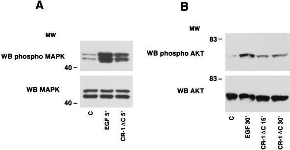
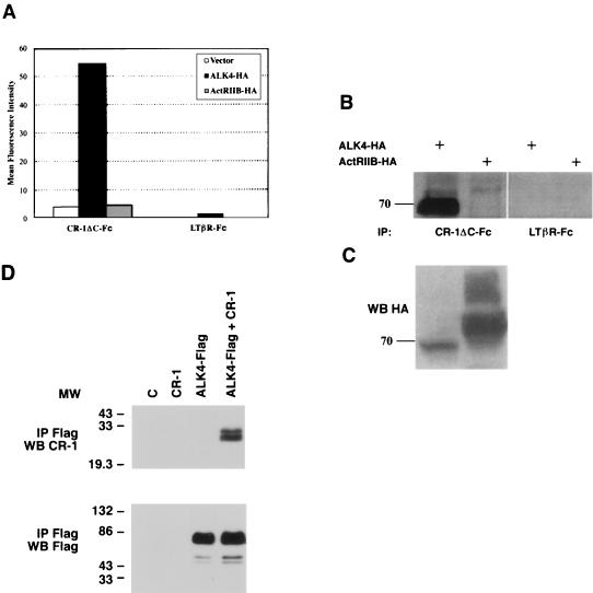
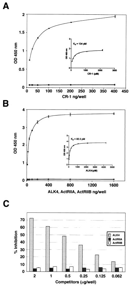
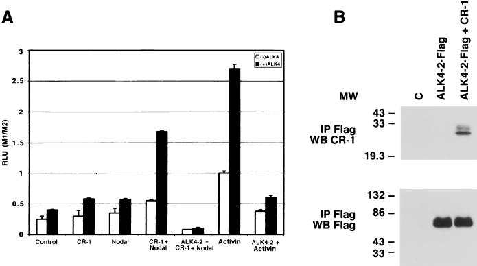
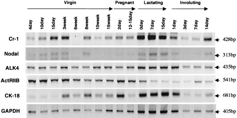
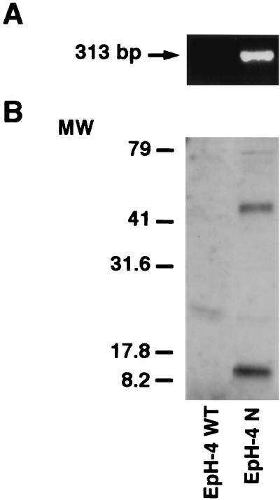
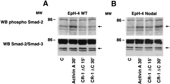
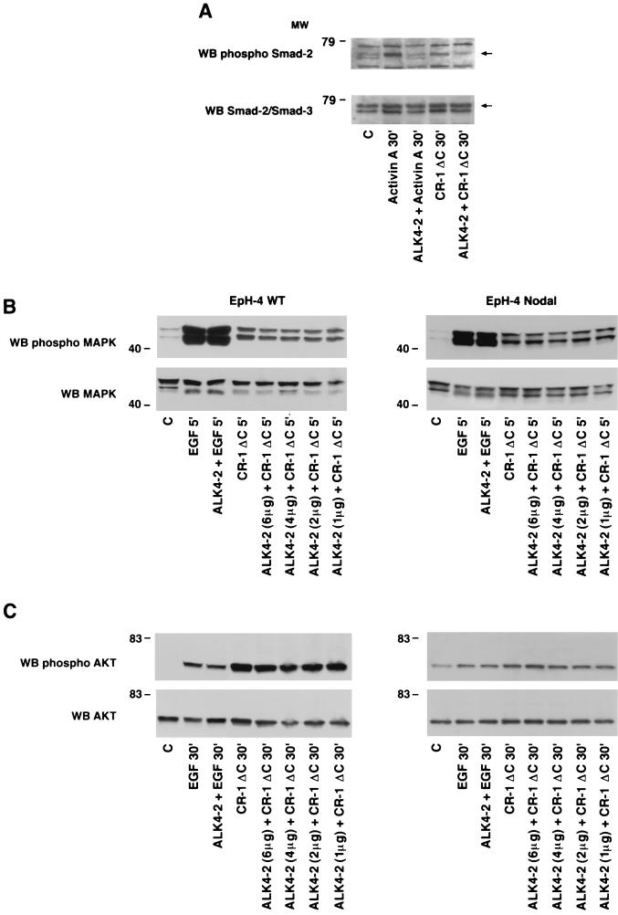
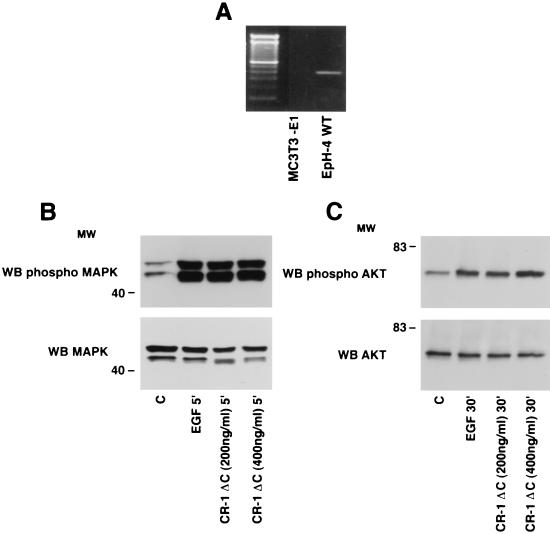
Cripto-1 (CR-1), an epidermal growth factor-CFC (EGF-CFC) family member, has a demonstrated role in embryogenesis and mammary gland development and is overexpressed in several human tumors. Recently, EGF-CFC proteins were implicated as essential signaling cofactors for Nodal, a transforming growth factor beta family member whose expression has previously been defined as embryo specific. To identify a receptor for CR-1, a human brain cDNA phage display library was screened using CR-1 protein as bait. Phage inserts with identity to ALK4, a type I serine/threonine kinase receptor for Activin, were identified. CR-1 binds to cell surface ALK4 expressed on mammalian epithelial cells in fluorescence-activated cell sorter analysis, as well as by coimmunoprecipitation. Nodal is coexpressed with mouse Cr-1 in the mammary gland, and CR-1 can phosphorylate the transcription factor Smad-2 in EpH-4 mammary epithelial cells only in the presence of Nodal and ALK4. In contrast, CR-1 stimulation of mitogen-activated protein kinase and AKT in these cells is independent of Nodal and ALK4, suggesting that CR-1 may modulate different signaling pathways to mediate its different functional roles.


Figures

FIG. 1.
FIG. 1.
MAPK and AKT activation by CR-1ΔC in EpH-4 cells. Serum-starved EpH-4 cells were stimulated with recombinant CR-1ΔC (200 ng/ml) or EGF (50 ng/ml) protein and analyzed by Western blot (WB) analysis. MW, molecular weight in thousands; C, control.
FIG. 2.
FIG. 2.
Binding of CR-1 to ALK4 expressed on the cell surface of mammalian epithelial cells. (A) 293 cells transiently transfected with ALK4-HA, ActRIIB-HA, or vector alone were incubated in the presence of CR-1ΔC-Fc recombinant protein or LTβR-Fc control protein. Binding of CR-1ΔC-Fc or LTβR-Fc protein to 293 cells was analyzed by FACS analysis as described in Materials and Methods. (B) 293 cells transiently transfected with ALK4-HA or ActRIIB-HA expression vector were immunoprecipitated (IP) with CR-1ΔC-Fc or LTβR-Fc and analyzed by Western blot analysis using an anti-HA rabbit polyclonal antibody. (C) As control, cell lysates from ALK4-HA- and ActRIIB-HA-transfected 293 cells were analyzed by Western blot analysis using an anti-HA rabbit polyclonal antibody. (D) COS1 cells were transiently transfected with ALK4-Flag and/or CR-1 expression vectors and Flag-tagged proteins were immunoprecipitated with an anti-Flag mouse monoclonal antibody. The immunoprecipitated proteins were analyzed by Western blot (WB) with an anti-CR-1 rabbit polyclonal antibody or an anti-Flag mouse monoclonal antibody. MW, molecular weight in thousands.
FIG. 3.
FIG. 3.
Binding of CR-1ΔC to the ECD of ALK4 (⧫), ActRIIA (▪), and ActRIIB (▵) in ELISA assay. (A) ECD of ALK4-Fc, ActRIIA-Fc, or ActRIIB-Fc (100 ng/well) was adsorbed to 96-well microtiter plates and incubated with various concentrations of CR-1ΔC recombinant protein (ng/100 μl). ELISA binding of soluble CR-1ΔC to immobilized proteins was detected as described in Materials and Methods. (B) CR-1ΔC (300 ng/well) was adsorbed to 96-well microtiter plates and incubated with various concentrations of the ECD of ALK4-Fc, ActRIIA-Fc, or ActRIIB-Fc recombinant protein (ng/100 μl). ELISA binding of soluble ECD of ALK4-Fc, ActRIIA, or ActRIIB to immobilized CR-1ΔC was detected as described in Materials and Methods. (C) One hundred nanograms of CR-1ΔC aloneor preincubated with different concentrations of ECD of ALK4-Fc, ActRIIA-Fc, or ActRIIB recombinant protein was added to plates that were preadsorbed with ECD of ALK4-Fc. ELISA binding of soluble CR-1ΔC to immobilized ECD of ALK4-Fc was detected as previously described. In panels A and B, the inserts show the Kd of each experiment. OD, optical density.
FIG. 4.
FIG. 4.
3TP dual-luciferase reporter assay in K562 cells transiently expressing ALK4-Flag, ALK4-2-Flag, CR-1, and/or Nodal. (A) Human K562 erythroleukemia cells were transfected with 0.5 μg of different plasmid DNAs together with Renilla luciferase control reporter vector (pRL-TK). Twenty-four hours after transfection, the cells were lysed and luciferase activity was measured using the Dual-Luciferase Reporter Assay system (Promega). M1/M2, plasmid DNA/control reporter vector ratio; RLU, relative light units. (B) Coimmunoprecipitation of dominant-negative ALK4-2-Flag with CR-1 in COS1 cells. COS1 cells transiently transfected with ALK4-2-Flag and/or CR-1 cDNAs were immunoprecipitated (IP) with an anti-Flag mouse monoclonal antibody and analyzed by Western blot (WB) using an anti-CR-1 rabbit polyclonal antibody or an anti-Flag mouse monoclonal antibody. MW, molecular weight in thousands; C, control.
FIG. 5.
FIG. 5.
RT-PCR for Cr-1, Nodal, ALK4, and ActRIIB in different stages of mouse mammary development. RT-PCR was performed using specific primers as described in Materials and Methods.
FIG. 6.
FIG. 6.
RT-PCR and Western blot analysis for Nodal in EpH-4 WT and Eph-4 N cells. (A) RT-PCR with specific Nodal primers (313 bp) in EpH-4 WT and EpH-4 N cells. (B) Western blot analysis using a rabbit polyclonal anti-Nodal antibody in the culture medium of EpH-4 WT and EpH-4 N cells. MW, molecular weight in thousands.
FIG. 7.
FIG. 7.
Smad-2 phosphorylation by CR-1ΔC in EpH-4 WT and EpH-4 N cells. Serum-starved EpH-4 WT (A) and EpH-4 N (B) cells were stimulated for different times with CR-1ΔC (400 ng/ml) or Activin A (50 ng/ml) recombinant protein and analyzed by Western blot (WB) analysis. MW, molecular weight in thousands; C, control.
FIG. 8.
FIG. 8.
Smad-2, MAPK, and AKT phosphorylation by CR-1ΔC in EpH-4 WT and EpH-4 N cells transiently transfected with a dominant-negative ALK4-2-Flag receptor. EpH-4 WT and EpH-4 N cells transiently transfected with ALK4-2-Flag or pCI-neo plasmid were stimulated with CR-1ΔC, EGF, or Activin A recombinant protein and cell lysates were assessed by Western blot (WB) analysis. (A) EpH-4 N cells transfected with pCI-neo (4 μg) (lanes 1, 2, and 4) and ALK4-2 (4 μg) were stimulated with Activin A (50 ng/ml) and CR-1ΔC (400 ng/ml) and analyzed by Western blot analysis. (B and C) EpH-4 WT or EpH-4 N cells transfected with pCI-neo (6 μg) (lanes 1, 2, and 4) and ALK4-2 (6, 4, 2, and 1 μg) were stimulated with EGF (50 ng/ml) and CR-1ΔC (200 ng/ml) and analyzed by Western blot analysis. MW, molecular weight in thousands; C, control.
FIG. 9.
FIG. 9.
MAPK and AKT phosphorylation by CR-1ΔC in MC3T3-E1 cells. (A) RT-PCR using specific primers for ALK4 (435 bp) in EpH-4 WT and MC3T3-E1 cells. A 100-bp DNA marker is shown in the first lane. (B and C) Serum-starved MC3T3-E1 cells were stimulated with CR-1ΔC (200 and 400 ng/ml) or EGF (50 ng/ml) recombinant protein and analyzed by Western blot (WB) analysis. MW, molecular weight in thousands; C, control.

References

    1. Attisano, L., C. Silvestri, L. Izzi, and E. Labbe. 2001. The transcriptional role of Smads and FAST (FoxH1) in TGFβ and activin signaling. Mol. Cell. Endocrinol. 180:3-11. - PubMed
    1. Bamford, R. N., E. Rossler, R. D. Burdine, U. Saplakoglu, J. dela Cruz, M. Splitt, J. Towbin, P. Bowers, B. Marino, A. F. Schier, M. M. Shen, M. M. Muenke, and B. Casey. 2000. Loss of function mutations in the EGF-CFC gene CFC1 are associated with human left-right laterality defects. Nat. Genet. 26:365-369. - PubMed
    1. Bianco, C., S. Kannan, M. De Santis, M. Seno, C. K. Tang, I. Martinez-Lacaci, N. Kim, B. Wallace-Jones, M. E. Lippman, A. Ebert, C. Wechselberger, and D. S. Salomon. 1999. Cripto-1 indirectly stimulates the tyrosine phosphorylation of erb B-4 through a novel receptor. J. Biol. Chem. 274:8624-8629. - PubMed
    1. Brandt, R., N. Normanno, W. J. Gullick, J. H. Lin, R. Harkins, D. Schneider, B. Jones, F. Ciardiello, M. G. Persico, F. Armenante, N. Kim, and D. S. Salomon. 1994. Identification and biological characterization of an epidermal growth factor-related protein: Cripto-1. J. Biol. Chem. 269:17320-17328. - PubMed
    1. Brennan, J., C. C. Lu, D. P. Norris, T. A. Rodriguez, R. S. Beddington, and E. J. Robertson. 2001. Nodal signalling in the epiblast patterns the early mouse embryo. Nature 411:965-969. - PubMed

Publication types

MeSH terms

Substances