. 2002 Apr;22(8):2743-50.
doi: 10.1128/MCB.22.8.2743-2750.2002.

Identification of mammalian Sds3 as an integral component of the Sin3/histone deacetylase corepressor complex

Affiliations

Identification of mammalian Sds3 as an integral component of the Sin3/histone deacetylase corepressor complex

Leila Alland et al. Mol Cell Biol. 2002 Apr.

Abstract

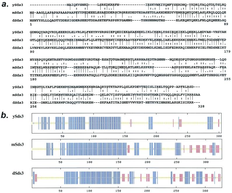
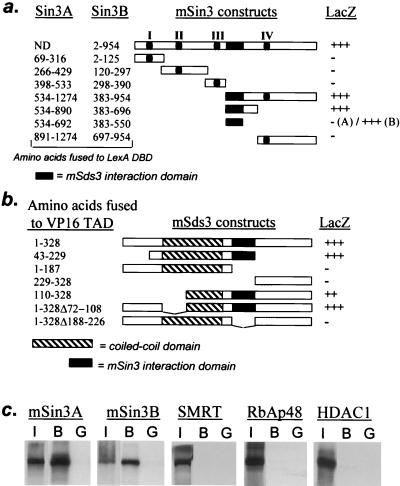
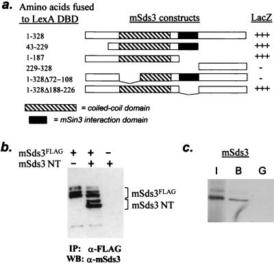
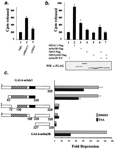
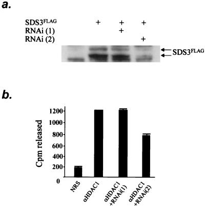
Silencing of gene transcription involves local chromatin modification achieved through the local recruitment of large multiprotein complexes containing histone deacetylase (HDAC) activity. The mammalian corepressors mSin3A and mSin3B have been shown to play a key role in this process by tethering HDACs 1 and 2 to promoter-bound transcription factors. Similar mechanisms appear to be operative in yeast, in which epistasis experiments have established that the mSin3 and HDAC orthologs (SIN3 and RPD3), along with a novel protein, SDS3, function in the same repressor pathway. Here, we report the identification of a component of the mSin3-HDAC complex that bears homology to yeast SDS3, physically associates with mSin3 proteins in vivo, represses transcription in a manner that is partially dependent on HDAC activity, and enables HDAC1 catalytic activity in vivo. That key physical and functional properties are also shared by yeast SDS3 underscores the central role of the Sin3-HDAC-Sds3 complex in eukaryotic cell biology, and the discovery of mSds3 in mammalian cells provides a new avenue for modulating the activity of this complex in human disease.


Figures

FIG. 1.
FIG. 1.
mSds3 encodes a mammalian protein that bears homology to yeast and Drosophila Sds3. (a) Amino acid comparisons of ySds3, mSds3, and dSds3. “|” indicates identity, “:” indicates high similarity, and “.” indicates low similarity. (b) Secondary structure predictions of ySds3, mSds3, and dSds3. Blue lines, alpha helix; red lines, beta sheet; yellow lines, indeterminate. Amino acids 59 to 170, 58 to 136, and 64 to 157 of mSds3, ySds3, and dSds3, respectively, correspond to predicted coiled-coil domains.
FIG. 2.
FIG. 2.
mSds3 interacts with mSin3A and mSin3B in yeast and in vitro. (a) Yeast two-hybrid studies using various mSin3A and mSin3B baits along with the VP16(TAD)-full-length mSds3 prey to localize mSin3's mSds3 interaction domain. I to IV correspond to the paired amphipatic helix of mSin3A and mSin3B (PAH). (b) Yeast two-hybrid studies using a full-length mSin3B bait along with various mSds3 prey to localize mSds3's mSin3 interaction domain. +++ indicates a LacZ phenotype that is equivalent to that observed for Myc-Max HLH/LZ interaction in yeast. (c) Bacterially expressed GST full-length mSds3 fusion protein was tested for its ability to interact with radiolabeled in vitro-translated mSin3A, mSin3B, or other known components of the mSin3 repression complex. I, input, 10%; B, bound (GST-mSds3); G, GST alone.
FIG. 3.
FIG. 3.
mSds3 interacts with mSin3A and mSin3B in vivo. (a and b) 293T cells were metabolically labeled after cotransfection of the various mSin3B and mSds3FLAG expression constructs shown. Radiolabeled whole-cell extracts were immunoprecipitated using anti-mSin3B antibody or anti-FLAG antibody and visualized by autoradiography. Triangles indicate the bands corresponding to the HID deletion mutant of mSin3B (panel b, lane 2) or the SID deletion mutant of mSds3 (panel b, lane 4). (c) Tissue extracts derived from various organs of newborn mice were immunoprecipitated with anti-mSds3, resulting in coimmunoprecipitation of mSin3A detected by Western blotting using mSin3A antibody (top panels), and similarly, extracts were immunoprecipitated with anti-mSin3A antibody, resulting in coimmunoprecipitation of mSds3 detected by Western blot using anti-mSds3 antibody (bottom panels). NRS, normal rabbit serum; HC, heavy chain.
FIG. 4.
FIG. 4.
mSds3 forms homodimers. (a) Yeast two-hybrid studies using various mSds3 baits along with VP16 TAD-full-length mSds3 prey to localize mSds3's dimerization domain. Helix A of the putative coiled-coil domain (residues 72 to 108) was defined by secondary structure prediction software. (b) In vivo expression of mSds3FLAG along with mSds3 N-terminal fragment (mSds3NT: residues 1 to 187) in 293T cells followed by immunoprecipitation with anti-FLAG antibody results in coimmunoprecipitation of mSds3NT detected by Western blot using anti-mSds3 antibody. IP, immunoprecipitation; WB, Western blot. (c) Bacterially expressed GST-full-length mSds3 fusion protein was tested for its ability to interact with radiolabeled in vitro translated full-length mSds3. I, input, 10%; B, bound (GST-mSds3); G, GST alone.
FIG. 5.
FIG. 5.
mSds3 forms a complex with mSin3B and HDAC1 in mammalian cells and mouse tissues. (a) Tissue extracts were derived as for Fig. 3c and immunoprecipitated with anti-HDAC1 antibody, resulting in coprecipitation of mSds3, detected by Western blot using mSds3 antibody. Anti-mSds3 immunoprecipitates were performed as a positive control for mSds3. NRS, normal rabbit serum; HC, heavy chain. (b) 293T cells were metabolically labeled after transfection of the various mSin3B, mSds3FLAG, and HDAC1 expression constructs shown. Whole-cell extracts were immunoprecipitated using anti-mSin3B antibody and visualized by autoradiography. The triangle indicates the band corresponding to the HID deletion mutants of mSin3B (lane 4). (c) NIH 3T3 cells were transfected with mSin3BFLAG and/or HDAC1FLAG expression constructs as shown. Whole-cell extracts were immunoprecipitated with an anti-mSds3 antibody, and the presence of mSin3BFLAG and/or HDAC1FLAG was detected by Western blotting using anti-FLAG antibody (right panel, lanes 4 to 6). The left panel (lanes 1 to 3) represents a Western blot performed with an anti-FLAG antibody on 10% of the extracts before immunoprecipitation.
FIG. 6.
FIG. 6.
mSds3 recruits histone deacetylase and represses basal transcription. (a and b) Deacetylase activity assays on immunoprecipitates from untransfected NIH 3T3 cells (a) or 293T cells transfected with the various expression constructs shown (b). Shown are the means from three experiments performed in duplicate and 95% confidence intervals. (a) endogenous histone deacetylase activity immunoprecipitated using normal rabbit serum (NRS) (lane 1), anti-HDAC1 antibody (lane 2), or anti-mSds3 antibody (lane 3). (b) Histone deacetylase assays on anti-FLAG immunoprecipitates from cells transfected with the indicated expression constructs (upper panel). The level of expression of transfected constructs was assessed by an anti-FLAG Western blot (bottom panel). (c) Reporter assays using various GAL4-mSds3 fusions and GAL4-mSin3B to repress a luciferase reporter gene containing GAL4 binding sites upstream of the myelomonocytic minimal promoter. The constructs and their levels of repression relative to GAL4 are shown. Shown are the means of three experiments performed in triplicate and 95% confidence intervals, except for GAL-mSin3B, for which the experiment was done once in triplicate. Gray bars, untreated cells; solid bars, cells treated with TSA for 24 h. DMSO, dimethyl sulfoxide.
FIG. 7.
FIG. 7.
mSds3 contributes to HDAC1 enzymatic activity. (a) Western blot performed with an anti-FLAG antibody on NIH 3T3 cell extracts after transfection with mSds3-FLAG construct with or without RNA primers. (b) Histone deacetylase activity assays were performed on anti-HDAC1 immunoprecipitates from NIH 3T3 cells after transfection with RNA primers. Shown are the means from three experiments. SDS3FLAG, mSds3FLAG.

References

    1. Alland, L., R. Muhle, H. Hou, Jr., J. Potes, L. Chin, N. Schreiber-Agus, and R. A. DePinho. 1997. Role for N-CoR and histone deacetylase in Sin3-mediated transcriptional repression. Nature 387:49-55. - PubMed
    1. Ayer, D. E., C. D. Laherty, Q. A. Lawrence, A. P. Armstrong, and R. N. Eisenman. 1996. Mad proteins contain a dominant transcription repression domain. Mol. Cell. Biol. 16:5772-5781. - PMC - PubMed
    1. Ayer, D. E., Q. A. Lawrence, and R. N. Eisenman. 1995. Mad-Max transcriptional repression is mediated by ternary complex formation with mammalian homologs of yeast repressor Sin3. Cell 80:767-776. - PubMed
    1. David, G., L. Alland, S. H. Hong, C. W. Wong, R. A. DePinho, and A. Dejean. 1998. Histone deacetylase associated with mSin3A mediates repression by the acute promyelocytic leukemia-associated PLZF protein. Oncogene 16:2549-2556. - PubMed
    1. Dhordain, P., O. Albagli, R. J. Lin, S. Ansieau, S. Quief, A. Leutz, J. P. Kerckaert, R. M. Evans, and D. Leprince. 1997. Corepressor SMRT binds the BTB/POZ repressing domain of the LAZ3/BCL6 oncoprotein. Proc. Natl. Acad. Sci. USA 94:10762-10767. - PMC - PubMed

Publication types

MeSH terms

Substances