. 2002 Apr;22(8):2777-87.
doi: 10.1128/MCB.22.8.2777-2787.2002.

Stat1-vitamin D receptor interactions antagonize 1,25-dihydroxyvitamin D transcriptional activity and enhance stat1-mediated transcription

Affiliations

Stat1-vitamin D receptor interactions antagonize 1,25-dihydroxyvitamin D transcriptional activity and enhance stat1-mediated transcription

Marcos Vidal et al. Mol Cell Biol. 2002 Apr.

Abstract

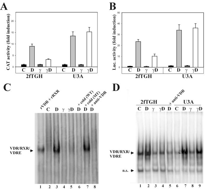
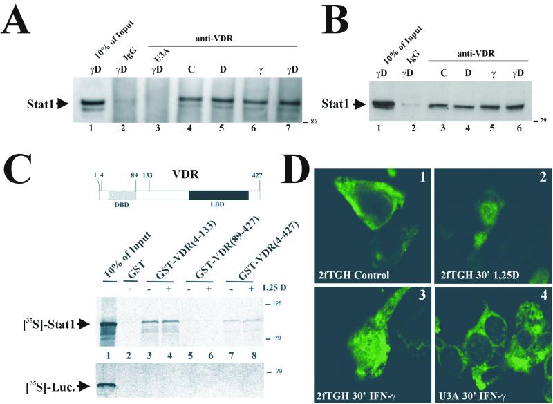
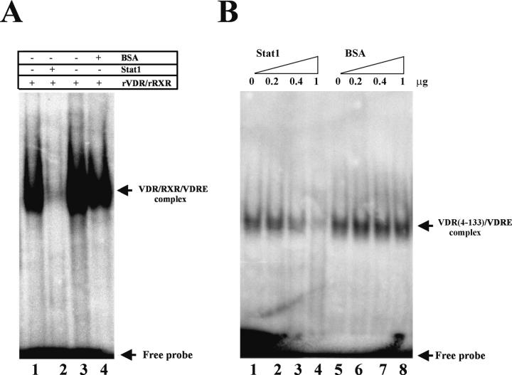
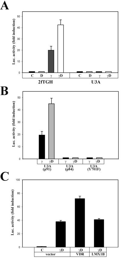
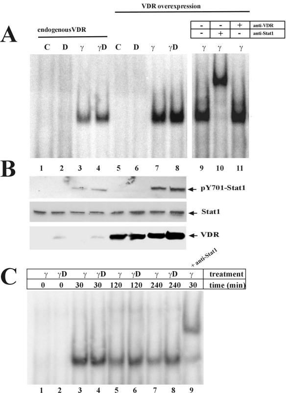
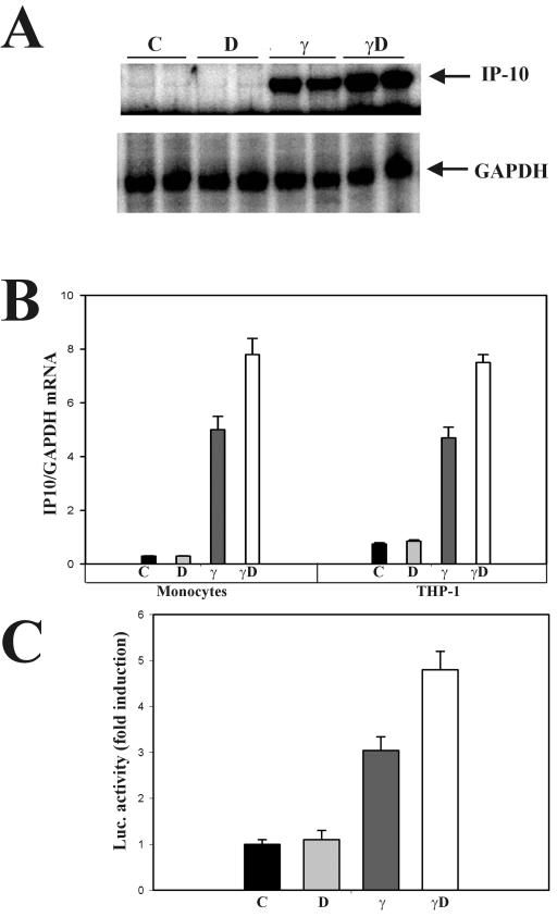
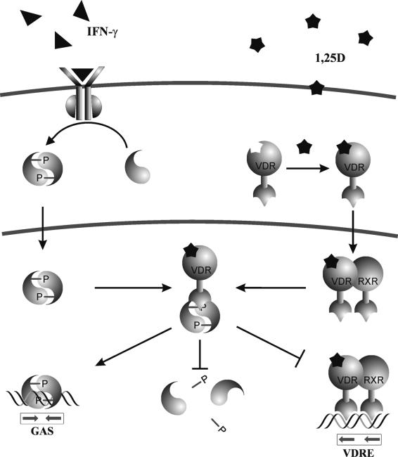
The cytokine gamma interferon (IFN-gamma) and the calcitropic steroid hormone 1,25-dihydroxyvitamin D (1,25D) are activators of macrophage immune function. In sarcoidosis, tuberculosis, and several granulomatoses, IFN-gamma induces 1,25D synthesis by macrophages and inhibits 1,25D induction of 24-hydroxylase, a key enzyme in 1,25D inactivation, causing high levels of 1,25D in serum and hypercalcemia. This study delineates IFN-gamma-1,25D cross talk in human monocytes-macrophages. Nuclear accumulation of Stat1 and vitamin D receptor (VDR) by IFN-gamma and 1,25D promotes protein-protein interactions between Stat1 and the DNA binding domain of the VDR. This prevents VDR-retinoid X receptor (RXR) binding to the vitamin D-responsive element, thus diverting the VDR from its normal genomic target on the 24-hydroxylase promoter and antagonizing 1,25D-VDR transactivation of this gene. In contrast, 1,25D enhances IFN-gamma action. Stat1-VDR interactions, by preventing Stat1 deactivation by tyrosine dephosphorylation, cooperate with IFN-gamma/Stat1-induced transcription. This novel 1,25D-IFN-gamma cross talk explains the pathogenesis of abnormal 1,25D homeostasis in granulomatous processes and provides new insights into 1,25D immunomodulatory properties.


Figures

FIG. 1.
FIG. 1.
IFN-γ antagonizes 1,25D transcriptional activity. (A) The human 24-hydroxylase promoter (−1262, +99) linked to CAT and a β-galactosidase expression plasmid were transiently transfected into wild-type 2fTGH or Stat1-null (U3A) cells. CAT activity was measured in cell lysates from untreated cells (C) or cells treated with 50 nM 1,25D (D), 1,000 U of IFN-γ/ml (γ), or both (γD) for 16 h. Results represent the means ± standard errors of the means of duplicate measurements from four independent experiments. (B) (VDRE)4-Luc and β-galactosidase expression plasmid were transiently transfected into 2fTGH or U3A cells. Luciferase activity was measured in cell lysates from cells treated as described for panel A. Results represent the means ± standard errors of the means of triplicate measurements from two independent experiments. (C) EMSA using the proximal VDRE of the human 24-hydroxylase promoter as a probe and nuclear extracts from THP-1 cells, treated for 4 h as described for panel A. Recombinant VDR and RXR (lane 1), incubation with a 300 M excess of cold wild-type (lane 6) or mutant (lane 7) VDRE, and incubation with anti-VDR 9A7 antibody (lane 8) served as controls. (D) EMSA using nuclear extracts from 2fTGH or U3A cells treated as described for panel C. n.s., nonspecific band.
FIG. 2.
FIG. 2.
VDR-Stat1 interaction. (A and B) Whole-cell extracts (600 μg of total protein) from VDR-transfected 2fTGH and U3A cells (A) or untransfected THP-1 (1 mg of total protein) (B) cells that were either untreated or treated with 50 nM 1,25D, 1,000 U of IFN-γ/ml, or both for 4 h were immunoprecipitated with anti-VDR 9A7 or nonspecific IgG and analyzed by Western blotting with anti-Stat1. (C) (Top) Schematic representation of the VDR. (Bottom) GST pull-down assay utilizing different GST-VDR fusion proteins purified from E. coli and bound to glutathione-agarose beads. Added was 1 μM 1,25D (+ lanes) or ethanol vehicle (− lanes). 35S-labeled Stat1 and luciferase were synthesized in vitro and incubated with the beads. After washing and elution, samples were subjected to SDS-10% polyacrylamide gel electrophoresis and autoradiographed. Numbers at right show molecular mass in kilodaltons. (D) Indirect VDR immunofluorescence in 2fTGH or U3A cells transiently transfected with VDR, either untreated or exposed to 50 nM 1,25D or 1,000 U of IFN-γ/ml for 30 min at 37°C, using a mouse monoclonal antibody against the VDR and detected with fluorescein isothiocyanate-conjugated anti-mouse antibody.
FIG. 3.
FIG. 3.
Stat1 abrogates VDR binding to VDRE. (A) EMSA using the proximal VDRE of the human 24-hydroxylase promoter as a probe and recombinant VDR and RXR (0.1 μg each) with or without recombinant Stat1 or BSA (1 μg each). Every reaction mixture contained 1 μM 1,25D. (B) EMSA performed as described for panel A using the N-terminal (amino acids 4 to 133) portion of the VDR (0.1 μg). Binding reaction mixtures included the indicated amounts (micrograms) of Stat1 or BSA.
FIG. 4.
FIG. 4.
1,25D synergy on IFN-γ-mediated transcription. (A) Cooperative effects of 1,25D on Stat1-induced transcription. (B) Requirements of the Stat1 molecule in 1,25D-IFN-γ synergy. (C) VDR overexpression enhances Stat1-mediated transcription. 2fTGH cells, U3A cells, and U3A cells reconstituted with different Stat1 mutants were transiently transfected with a luciferase reporter construct driven by eight copies of the consensus GAS (TTCTCGGAA) and 0.1 μg of either human VDR expression vector, human LMX1B expression vector, or vector alone when indicated. Twenty-four hours after transfection, cells were either untreated (C) or exposed to 10 nM 1,25D (D), 1,000 IU of IFN-γ/ml (γ), or both (γD) as indicated for 4 h. Luciferase activity was determined in cell lysates. Bars and error bars represent means ± standard errors of the means from triplicate measurements from two (C) or three (A and B) independent experiments.
FIG. 5.
FIG. 5.
VDR prolongs Stat1 activation. (A) EMSA using a consensus GAS as a probe and nuclear extracts from untransfected 2fTGH cells or from cells transiently transfected with human VDR expression vector. Cells were either untreated or treated for 4 h with 50 nM 1,25D, 1,000 U of IFN-γ/ml, or both. The right panel shows supershift analysis utilizing anti-VDR or anti-Stat1 antibodies. (B) Western blots from cell extracts treated as described for panel A, probed with anti-pY701 Stat1, anti-Stat1, and anti-VDR. (C) EMSA analysis was done as described for panel A with cell extracts from untransfected 2fTGH cells that were treated with 1,000 U of IFN-γ/ml or 1,000 U of IFN-γ/ml plus 50 nM 1,25D for the indicated periods of time.
FIG. 6.
FIG. 6.
1,25D synergy on IFN-γ induction of the IP-10 gene. (A) Normal blood monocytes were left untreated or treated with 50 nM 1,25D, 150 U of IFN-γ/ml, or both for 18 h. IP-10 and GAPDH mRNAs were measured by RNase protection assays. (B) Relative IP-10/GAPDH mRNA levels in peripheral blood monocytes from four healthy volunteers (left graph) or differentiated THP-1 cells (right graph) from two independent experiments. Results represent the means ± standard errors of the means. (C) 2fTGH cells were transiently transfected with TGL-IP10. Twenty-four hours after transfection, cells were either untreated (C) or exposed to 10 nM 1,25D (D), 500 IU of IFN-γ/ml (γ), or both (γD) as indicated for 24 h. Luciferase activity was determined in cell lysates. Bars and error bars represent means ± standard errors of the means from triplicate measurements.
FIG. 7.
FIG. 7.
Schematic diagram of functional Stat1-VDR interactions. Nuclear accumulation of Stat1 and the VDR by IFN-γ and 1,25D promotes physical interaction between Stat1 and the DBD of the VDR. Stat1-VDR complex formation inhibits VDR-RXR binding to VDRE and Stat1 deactivation by tyrosine dephosphorylation, thus resulting in IFN-γ antagonism on 1,25D-VDR transactivation and 1,25D cooperation on Stat1-mediated transcription.

References

    1. Adams, J. S., M. A. Gacad, A. Anders, D. B. Endres, and O. P. Sharma. 1986. Biochemical indicators of disordered vitamin D and calcium homeostasis in sarcoidosis. Sarcoidosis 3:1-6. - PubMed
    1. Adams, J. S., R. L. Modlin, M. M. Diz, and P. F. Barnes. 1989. Potentiation of the macrophage 25-hydroxyvitamin D-1-hydroxylation reaction by human tuberculous pleural effusion fluid. J. Clin. Endocrinol. Metab. 69:457-460. - PubMed
    1. Adams, J. S., F. R. Singer, M. A. Gacad, O. P. Sharma, M. J. Hayes, P. Vouros, and M. F. Holick. 1985. Isolation and structural identification of 1,25-dihydroxyvitamin D3 produced by cultured alveolar macrophages in sarcoidosis. J. Clin. Endocrinol. Metab. 60:960-966. - PubMed
    1. Aittomaki, S., M. Pesu, B. Groner, O. A. Janne, J. J. Palvimo, and O. Silvennoinen. 2000. Cooperation among Stat1, glucocorticoid receptor, and PU.1 in transcriptional activation of the high-affinity Fc gamma receptor I in monocytes. J. Immunol. 164:5689-5697. - PubMed
    1. Alroy, I., T. L. Towers, and L. P. Freedman. 1995. Transcriptional repression of the interleukin-2 gene by vitamin D3: direct inhibition of NFATp/AP-1 complex formation by a nuclear hormone receptor. Mol. Cell. Biol. 15:5789-5799. - PMC - PubMed

Publication types

MeSH terms

LinkOut - more resources