. 2002 Apr;22(8):2821-9.
doi: 10.1128/MCB.22.8.2821-2829.2002.

Impaired cardiac contractility response to hemodynamic stress in S100A1-deficient mice

Affiliations

Impaired cardiac contractility response to hemodynamic stress in S100A1-deficient mice

Xiao-Jun Du et al. Mol Cell Biol. 2002 Apr.

Abstract

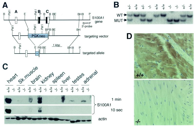
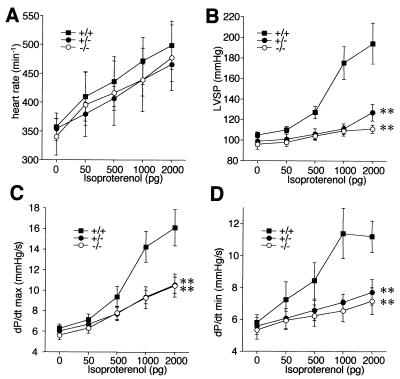
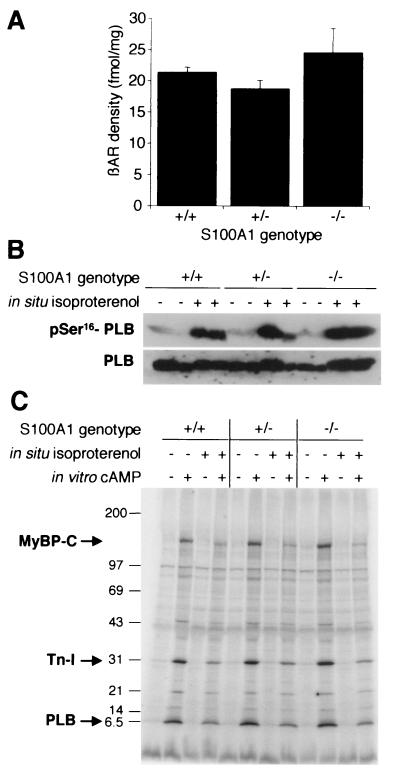
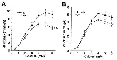
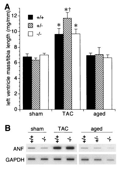
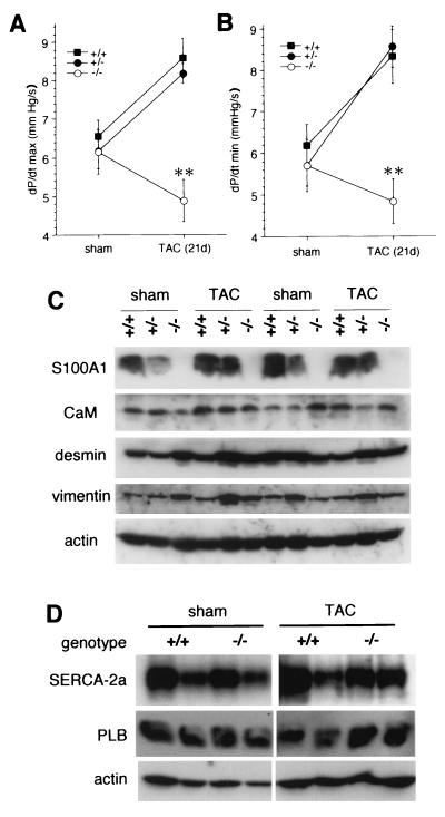
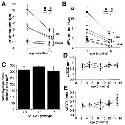
Ca(2+) signaling plays a central role in cardiac contractility and adaptation to increased hemodynamic demand. We have generated mice with a targeted deletion of the S100A1 gene coding for the major cardiac isoform of the large multigenic S100 family of EF hand Ca(2+)-binding proteins. S100A1(-/-) mice have normal cardiac function under baseline conditions but have significantly reduced contraction rate and relaxation rate responses to beta-adrenergic stimulation that are associated with a reduced Ca(2+) sensitivity. In S100A1(-/-) mice, basal left-ventricular contractility deteriorated following 3-week pressure overload by thoracic aorta constriction despite a normal adaptive hypertrophy. Surprisingly, heterozygotes also had an impaired response to acute beta-adrenergic stimulation but maintained normal contractility in response to chronic pressure overload that coincided with S100A1 upregulation to wild-type levels. In contrast to other genetic models with impaired cardiac contractility, loss of S100A1 did not lead to cardiac hypertrophy or dilation in aged mice. The data demonstrate that high S100A1 protein levels are essential for the cardiac reserve and adaptation to acute and chronic hemodynamic stress in vivo.


Figures

FIG. 1.
FIG. 1.
Generation and analysis of S100A1-targeted mice. (A) Map of the murine S100A1 gene, targeting vector, and targeted allele after Cre/LoxP recombination. Boxes and bold letters indicate exons, with protein-coding sequences shown in black. Selected restriction enzyme cleavage sites are indicated: B, BglII; BH, BamHI; E, EcoRI; P, PvuII. ×, loxP. (B) Southern blot genotyping of a litter of an S100A1 heterozygous cross. WT, wild-type allele; MUT, mutant allele. (C) Immunoblot analysis of tissue extracts from S100A1−/− mice and wild-type littermates. Two S100A1 exposures and an actin loading control are shown. (D) Immunohistochemistry of wild-type and mutant left ventricles. The localization of S100A1 is indicated by the brown color.
FIG. 2.
FIG. 2.
β-Adrenergic cardiac response in intact animals measured with a microcatheter placed in the left ventricle. (A) Heart rates; (B) left-ventricular systolic pressure (LVSP); (C) maximal contraction rates (dP/dtmax, in thousands); (D) maximal relaxation rates (dP/dtmin, in thousands). Isoproterenol was intravenously given as a bolus as indicated. ∗∗, P < 0.001 versus the wild type.
FIG. 3.
FIG. 3.
Normal β-adrenergic pathway in S100A1-deficient mice. (A) β-Adrenoceptor (βAR) densities in left-ventricle membrane protein preparations. (B) Isoproterenol-induced phosphorylation of PLB. Results from two animals per genotype are shown. Hearts were perfused in situ with or without 1 μM isoproterenol, and protein extracts of left ventricles were analyzed with a phospho-Ser16-specific antibody. The lower panel shows a similarly loaded blot analyzed for total PLB as a loading control. (C) Autoradiograph of a representative back-phosphorylation assay. Left-ventricle protein extracts from control (−) or isoproterenol-stimulated (+) hearts were subjected to in vitro phosphorylation assays with or without cAMP addition and separated by SDS-5 to 18% polyacrylamide gel electrophoresis. Positions of mass standards are indicated on the left (in kilodaltons). MyBP-C, myosin-binding protein C; Tn-I, troponin I.
FIG. 4.
FIG. 4.
Inotropic (A) and lusitropic (B) responses in hearts perfused in situ with various Ca2+ concentrations. ∗∗, P < 0.001 versus wild type. n = 7 per genotype; values are in thousands.
FIG. 5.
FIG. 5.
Left-ventricular hypertrophy indicators. (A) Wet weights of left ventricles normalized by tibia length from 5-month-old (sham and TAC) and 16-month-old (aged) mice. (B) RNA blot analysis of left ventricles using ANF and GAPDH cDNA probes. ∗, P < 0.01 versus the respective sham-operated group; †, P < 0.05 versus the −/− TAC-treated group.
FIG. 6.
FIG. 6.
Functional response to chronic hemodynamic stress induced by 3-week TAC compared to sham-operated littermates. (A and B) Basal left-ventricular contractility (A) and relaxation (B) rates were measured in microcatheterized intact animals. Values are in thousands. (C) Immunoblot analysis of heart extracts of sham-operated and TAC-treated mice, probed with the antibodies indicated on the left. CaM, calmodulin. (D) SERCA-2a and PLB levels in sham- and TAC-treated wild-type and S100A1 null mice. ∗∗, P < 0.001 versus wild type and heterozygotes.
FIG. 7.
FIG. 7.
Cardiac parameters in aged mice. (A and B) Basal and isoproterenol (2 ng)-stimulated inotropic (A) rates and lusitropic (B) rates in 16-month-old versus 5-month-old mice. Values are in thousands. (C) Left-ventricular cardiomyocyte cross-sectional areas in 16-month-old mice. (D) Left-ventricle end diastolic diameters (LVEDD) in groups of 10 mice per genotype from 10 weeks to 16 months of age. (E) Anterior wall diastolic thicknesses (AWDTh) in the same groups of animals shown in panel D. Posterior wall thicknesses were similar to those shown here (data not shown). ∗, P < 0.05 versus wild type.

References

    1. Arber, S., J. J. Hunter, J. Ross, M. Hongo, G. Sansig, J. Borg, J. C. Perriard, K. R. Chien, and P. Caroni. 1997. MLP-deficient mice exhibit a disruption of cardiac cytoarchitectural organization, dilated cardiomyopathy, and heart failure. Cell 88:393-403. - PubMed
    1. Baudier, J., E. Bergeret, N. Bertacchi, H. Weintraub, J. Gagnon, and J. Garin. 1995. Interactions of myogenic bHLH transcription factors with calcium-binding calmodulin and S100a (aa) proteins. Biochemistry 34:7834-7846. - PubMed
    1. Chawengsaksophak, K., R. James, V. E. Hammond, F. Köntgen, and F. Beck. 1997. Homeosis and intestinal tumours in Cdx2 mutant mice. Nature 386:84-87. - PubMed
    1. Chien, K. R. 2000. Genomic circuits and the integrative biology of cardiac diseases. Nature 407:227-232. - PubMed
    1. Clapham, D. E. 1995. Calcium signalling. Cell 80:259-268. - PubMed

Publication types

MeSH terms