. 2002 Apr;22(8):2862-70.
doi: 10.1128/MCB.22.8.2862-2870.2002.

NF-kappa B activates prostate-specific antigen expression and is upregulated in androgen-independent prostate cancer

Affiliations

NF-kappa B activates prostate-specific antigen expression and is upregulated in androgen-independent prostate cancer

Charlie D Chen et al. Mol Cell Biol. 2002 Apr.

Abstract

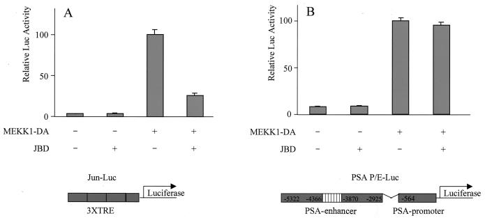
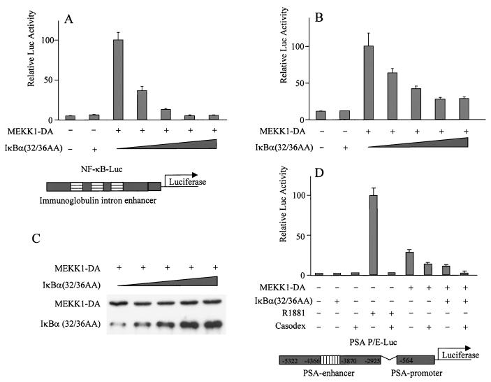
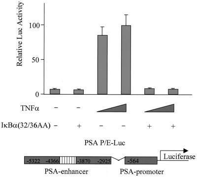
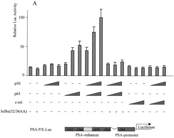
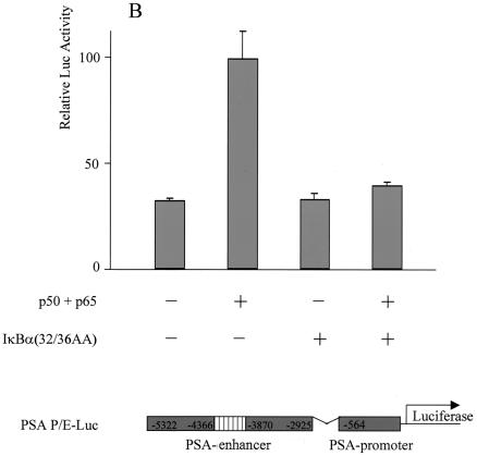
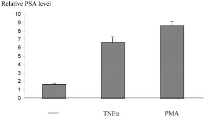
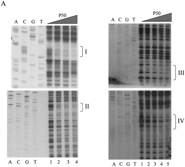
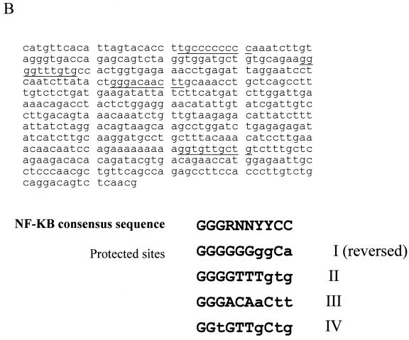
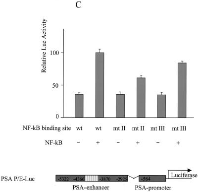
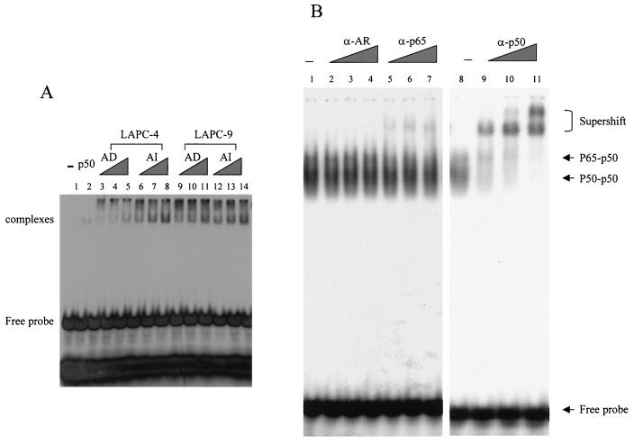
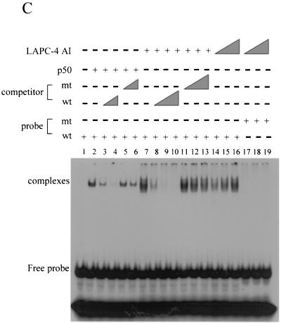
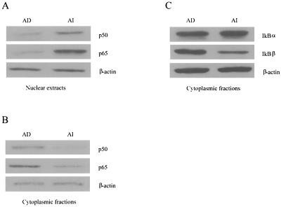
The transcription factor NF-kappa B regulates gene expression involved in cell growth and survival and has been implicated in progression of hormone-independent breast cancer. By expressing a dominant-active form of mitogen-activated protein kinase kinase kinase 1, by exposure to tumor necrosis factor alpha, or by overexpression of p50/p65, we show that NF-kappa B activates a transcription regulatory element of the prostate-specific antigen (PSA)-encoding gene, a marker for prostate cancer development, treatment, and progression. By DNase I footprinting, we identified four NF-kappa B binding sites in the PSA core enhancer. We also demonstrate that androgen-independent prostate cancer xenografts have higher constitutive NF-kappa B binding activity than their androgen-dependent counterparts. These results suggest a role of NF-kappa B in prostate cancer progression.


Figures

FIG. 1.
FIG. 1.
JBD does not inhibit MEKK1-mediated activation of PSA. LNCaP cells were transfected with empty vector pcDNA3, JBD (500 ng), MEKK1-DA (400 ng), or JBD with MEKK1-DA, together with the reporter construct driven either by a Jun-responsive element (Jun-Luc; 200 ng) (A) or by PSA-P/E-Luc (200 ng) (B). Luciferase activity was measured 2 days after transfection and normalized by transfection efficiency, which was determined by GFP cotransfection. The data shown represent three experiments.
FIG. 2.
FIG. 2.
Dominant-active IκBα mutant inhibits MEKK1-mediated activation of PSA. LNCaP cells were transfected with empty vector pcDNA3, the mutant form IκBα(32/36AA) (800 ng), MEKK1-DA (300 ng), or MEKK1-DA with increasing amounts (100, 200, 400, and 800 ng) of the mutant form IκBα(32/36AA), together with the reporter construct driven either by an NF-κB-responsive element (NF-κB-Luc; 200 ng) (A) or by PSA-P/E-Luc (200 ng) (B and D). (C) Lysates from the transfected cells were examined for MEKK1-DA expression. Bicalutamide was used as indicated in the experiment whose results are shown in panel D. Luciferase activity was measured 2 days after transfection and normalized by transfection efficiency, which was determined by GFP cotransfection. The data shown represent three experiments.
FIG. 3.
FIG. 3.
TNF-α activates PSA expression through the NF-κB pathway. LNCaP cells were transfected with the reporter construct PSA-P/E-Luc (200 ng) with or without the mutant form IκBα(32/36AA) (800 ng) as indicated. After transfection, cells were treated with or without TNF-α (10 or 50 ng/ml) as indicated. Luciferase activity was measured 2 days after transfection and normalized to Renilla luciferase. The data shown represent two experiments.
FIG. 4.
FIG. 4.
Overexpression of NF-κB activated PSA expression, and the activation was abolished by mutant IκBα. (A) LNCaP cells were transfected with the reporter construct PSA-P/E-Luc (200 ng) with increasing amounts of human p50 (p50) (25, 50, and 100 ng), human p65 (p65) (25, 50, and 100 ng), a combination of p50 and p65, c-Rel (25, 50, and 100 ng), or a combination of p50 and c-Rel and with or without the mutant form IκBα(32/36AA) (800 ng) as indicated. (B) DU145 cells were cotransfected with the reporter construct PSA-P/C-Luc (200 ng) and p50 (50 ng) and p65 (50 ng) and with or without the mutant form IκBα(32/36AA) (800 ng) as indicated. Luciferase activity was measured 2 days after transfection and normalized to Renilla luciferase. The data shown represent two experiments.
FIG. 4.
FIG. 4.
Overexpression of NF-κB activated PSA expression, and the activation was abolished by mutant IκBα. (A) LNCaP cells were transfected with the reporter construct PSA-P/E-Luc (200 ng) with increasing amounts of human p50 (p50) (25, 50, and 100 ng), human p65 (p65) (25, 50, and 100 ng), a combination of p50 and p65, c-Rel (25, 50, and 100 ng), or a combination of p50 and c-Rel and with or without the mutant form IκBα(32/36AA) (800 ng) as indicated. (B) DU145 cells were cotransfected with the reporter construct PSA-P/C-Luc (200 ng) and p50 (50 ng) and p65 (50 ng) and with or without the mutant form IκBα(32/36AA) (800 ng) as indicated. Luciferase activity was measured 2 days after transfection and normalized to Renilla luciferase. The data shown represent two experiments.
FIG. 5.
FIG. 5.
TNF-α and PMA induce endogenous PSA expression. Serum-starved LNCaP cells were grown in 5% charcoal-stripped FBS with or without TNF-α (50 nM) or PMA (10 ng/ml) overnight, and the media were subjected to ELISA to determine PSA expression.
FIG. 6.
FIG. 6.
Identification of NF-κB binding sites in PSA core enhancer. (A) Increasing amounts of recombinant human p50 protein (0, 0.5, 1.0, 2.0, and 4.0 gel shift units in lanes 1, 2, 3, 4, and 5, respectively) were incubated with a 32P-labeled enhancer fragment, from −4366 to −3824, to detect footprints. The sites identified are designated I, II, III, and IV in sequence order. Sequencing ladders are included alongside the footprints to localize the protected sites. The order of binding affinity was I > II > III > IV. (B) Sequence of the PSA core enhancer. Footprinted regions are underlined and compiled below the consensus. The sequence of site I is reversed. (C) Wild-type (wt) or mutant PSA-P/E-Luc was cotransfected with p50 and p65 into LNCaP cells. Luciferase activity was measured 2 days after transfection and normalized by transfection efficiency, which was determined by GFP cotransfection. The data shown represent three experiments. Sites II and III were mutated to CCGGTTTGTG and ACGGAGTACT, respectively.
FIG. 6.
FIG. 6.
Identification of NF-κB binding sites in PSA core enhancer. (A) Increasing amounts of recombinant human p50 protein (0, 0.5, 1.0, 2.0, and 4.0 gel shift units in lanes 1, 2, 3, 4, and 5, respectively) were incubated with a 32P-labeled enhancer fragment, from −4366 to −3824, to detect footprints. The sites identified are designated I, II, III, and IV in sequence order. Sequencing ladders are included alongside the footprints to localize the protected sites. The order of binding affinity was I > II > III > IV. (B) Sequence of the PSA core enhancer. Footprinted regions are underlined and compiled below the consensus. The sequence of site I is reversed. (C) Wild-type (wt) or mutant PSA-P/E-Luc was cotransfected with p50 and p65 into LNCaP cells. Luciferase activity was measured 2 days after transfection and normalized by transfection efficiency, which was determined by GFP cotransfection. The data shown represent three experiments. Sites II and III were mutated to CCGGTTTGTG and ACGGAGTACT, respectively.
FIG. 7.
FIG. 7.
AI tumors have a higher level of constitutive NF-κB binding activity than do AD tumors. (A) Increasing amounts (0.5, 1.0, and 2.0 μg) of nuclear extracts from both AD and AI LAPC-4 and LAPC-9 tumors were incubated with a 32P-labeled NF-κB binding sequence to detect NF-κB binding by gel shift analysis. The shifted complex and free probe are indicated. (B) Identity of the shifted complexes. Two micrograms of LAPC-4 AI tumor nuclear extract was incubated with a 32P-labeled NF-κB binding sequence with or without increasing amounts of antibodies against AR, p65, or p50. (C) Specificity of the shifted complexes. Shifted complexes (lane 2) formed by the recombinant p50 protein were competed away by increasing amounts of the unlabeled wild-type (wt) probe (lanes 3 and 4) but not by the unlabeled mutant (mt) probe (lanes 5 and 6). LAPC-4 AI tumor nuclear extracts (0.5, 1.0, and 2.0 μg) formed a band shift with the wild-type probe (lanes 14 to 16) and but not with the mutant probe (lanes 17 to 19). The band shift was competed away by increasing amounts of the unlabeled wild-type probe (lanes 8 to 10) but not by the unlabeled mutant probe (lanes 11 to 13).
FIG. 7.
FIG. 7.
AI tumors have a higher level of constitutive NF-κB binding activity than do AD tumors. (A) Increasing amounts (0.5, 1.0, and 2.0 μg) of nuclear extracts from both AD and AI LAPC-4 and LAPC-9 tumors were incubated with a 32P-labeled NF-κB binding sequence to detect NF-κB binding by gel shift analysis. The shifted complex and free probe are indicated. (B) Identity of the shifted complexes. Two micrograms of LAPC-4 AI tumor nuclear extract was incubated with a 32P-labeled NF-κB binding sequence with or without increasing amounts of antibodies against AR, p65, or p50. (C) Specificity of the shifted complexes. Shifted complexes (lane 2) formed by the recombinant p50 protein were competed away by increasing amounts of the unlabeled wild-type (wt) probe (lanes 3 and 4) but not by the unlabeled mutant (mt) probe (lanes 5 and 6). LAPC-4 AI tumor nuclear extracts (0.5, 1.0, and 2.0 μg) formed a band shift with the wild-type probe (lanes 14 to 16) and but not with the mutant probe (lanes 17 to 19). The band shift was competed away by increasing amounts of the unlabeled wild-type probe (lanes 8 to 10) but not by the unlabeled mutant probe (lanes 11 to 13).
FIG. 8.
FIG. 8.
Western blot analysis of NF-κB and IκB protein levels in LAPC-4 xenograft tumors. (A) Nuclear extracts were subjected to Western blot analysis with antibodies against p50 and p65. (B) Cytoplasmic fractions were subjected to Western blot analysis with antibodies against p50 and p65. (C) Cytoplasmic fractions were subjected to Western blot analysis with antibodies against IκBα and IκBβ.

References

    1. Aarnisalo, P., H. Santti, H. Poukka, J. J. Palvimo, and O. A. Janne. 1999. Transcription activating and repressing functions of the androgen receptor are differentially influenced by mutations in the deoxyribonucleic acid binding domain. Endocrinology 140:3097-3105. - PubMed
    1. Abreu-Martin, M. T., A. Chari, A. A. Palladino, N. A. Craft, and C. L. Sawyers. 1999. Mitogen-activated protein kinase kinase kinase 1 activates androgen receptor-dependent transcription and apoptosis in prostate cancer. Mol. Cell. Biol. 19:5143-5154. - PMC - PubMed
    1. Baldwin, A. S., Jr. 2001. Series introduction: the transcription factor NF-κB and human disease. J. Clin. Investig. 107:3-6. - PMC - PubMed
    1. Beg, A. A., W. C. Sha, R. T. Bronson, S. Ghosh, and D. Baltimore. 1995. Embryonic lethality and liver degeneration in mice lacking the RelA component of NF-κB. Nature 376:167-170. - PubMed
    1. Chadwick, C. C., L. J. Shaw, and R. C. Winneker. 1998. TNF-α and 9-cis-retinoic acid synergistically induce ICAM-1 expression: evidence for interaction of retinoid receptors with NF-κB. Exp. Cell Res. 239:423-429. - PubMed

Publication types

LinkOut - more resources