. 2002 Apr;22(8):2871-81.
doi: 10.1128/MCB.22.8.2871-2881.2002.

MSK1 and MSK2 are required for the mitogen- and stress-induced phosphorylation of CREB and ATF1 in fibroblasts

Affiliations

MSK1 and MSK2 are required for the mitogen- and stress-induced phosphorylation of CREB and ATF1 in fibroblasts

Giselle R Wiggin et al. Mol Cell Biol. 2002 Apr.

Abstract

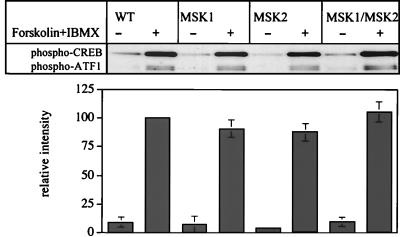
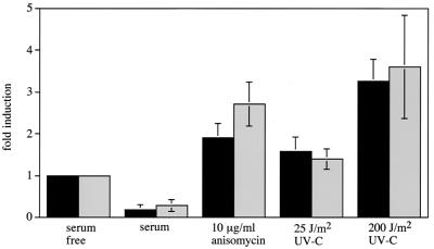
Using mouse knockouts for mitogen- and stress-activated protein kinase 1 (MSK1) and MSK2 and a double knockout of both MSK1 and MSK2, we show that these protein kinases are required for the stress-induced phosphorylation of transcription factors CREB and ATF1 in primary embryonic fibroblasts. In contrast mitogen-induced phosphorylation of CREB and ATF1 is greatly reduced but not totally abolished. The mitogen- and stress-induced phosphorylation of CREB at Ser133 has been linked to the transcription of several immediate early genes, including c-fos, junB, and egr1. The knockout of both MSK1 and MSK2 resulted in a 50% reduction in c-fos and junB gene transcription in response to anisomycin or UV-C radiation but only a small reduction in response to tetradecanoyl phorbol acetate or epidermal growth factor in fibroblasts. The transcription of egr1 in response to both mitogenic and stress stimuli, as well as stress-induced apoptosis, was unaffected in the MSK1/MSK2 double knockout.


Figures

FIG. 1.
FIG. 1.
Generation of ES cells with a targeted MSK2 gene. (A) A targeting vector was made to delete exons 5 to 7 of the murine MSK2 gene through the addition of a neomycin selection cassette and polyadenylation sequences. A thymidine kinase cassette acts as a negative selection marker. Positions of the 3′ and 5′ probes used to screen for the correct incorporation of the targeting vector by NcoI digestion are shown. (B and C) Genomic DNA from wild-type and MSK2 heterozygous ES cells was digested with NcoI, run on an 0.8% agarose gel, and Southern blotted with either the 3′ (B) or 5′ (C) probe. An 11.8-kb band indicating a wild-type locus and an 8.4-kb band indicating a targeted locus (due to an additional NcoI site within the neomycin selection cassette) are shown for blots with the 3′ probe. For blots with the 5′ probe, an 11.8-kb band for the wild-type locus and a 3.4-kb band for a targeted locus are shown.
FIG. 2.
FIG. 2.
Genotyping and expression levels of MSK1 and MSK2 in the knockout mice. (A) Genotyping for MSK1 (top) and MSK2 (bottom) knockout mice from a tail biopsy sample. DNA samples were subjected to PCR as described in Materials and Methods, electrophoresed on 1.5% (wt/vol) agarose gels, and examined by ethidium bromide staining. (B) Lysates from wild-type, MSK1, MSK2, and MSK1/MSK2 knockout fibroblasts (30 μg of protein) were immunoblotted as described in Materials and Methods with an antibody raised against the MSK1 protein. Alternatively, MSK2 was immunoprecipitated from 1 mg of fibroblast cell lysate protein using an antipeptide antibody raised against residues 753 to 772 of MSK2 in sheep. The immunoprecipitates were resuspended in SDS, electrophoresed, and immunoblotted as described in Materials and Methods with an antibody against the MSK2 protein raised in rabbits. Lysates from two different dishes of cells were used for each condition.
FIG.3.
FIG.3.
Inhibition of MAPK cascades by PD-184352 and SB-203580 in wild-type fibroblasts. Fibroblasts were serum starved overnight, preincubated for 1 h with or without PD-184352 (5 μM), SB-203580 (10 μM), or both PD-184352 and SB-203580 and then incubated with or without TPA (400 ng/ml, 10 min) or EGF (100 ng/ml, 5 min) or exposed to UV-C radiation (200 J/m2, followed by a 15-min incubation at 37°C) or anisomycin (10 μg/ml, 30 min). The cells were then lysed. (A) Cell lysates were immunoblotted with antibodies that recognize ERK1 and ERK2 or SAPK2/p38 only when they are phosphorylated at the Thr-X-Tyr motif. (B) MAPKAP-K1b/RSK2, MAPKAP-K2, MSK1, and MSK2 were immunoprecipitated from fibroblast lysates and assayed. Further details are given in Materials and Methods. One unit of activity was that amount of enzyme that incorporates 1 nmol of phosphate into the peptide substrate in 1 min. Error bars represent the standard errors of the means of duplicate immunoprecipitations from three different dishes of cells.
FIG. 4.
FIG. 4.
Activation of components of MAPK cascades from different MSK knockout fibroblasts. (A) Confluent fibroblasts from wild-type (WT), MSK1 knockout, MSK2 knockout, and MSK1/MSK2 double-knockout embryos were serum starved overnight and then left unstimulated (−) or stimulated (+) with TPA (400 ng/ml, 10 min), EGF (100 ng/ml, 5 min), anisomycin (10 μg/ml, 30 min), or UV-C (200 J/m2, followed by a 15-min incubation at 37°C). Cells were then lysed, and 20 μg of lysate protein was denatured in SDS, electrophoresed on 4 to 12% Novex polyacrylamide gels, transferred to nitrocellulose membranes, and immunoblotted for phosphorylated ERK1 and ERK2 or for phosphorylated SAPK2/p38. The levels of ERK1 and ERK2 and SAPK2/p38 were measured with an antibody that recognizes the phosphorylated and dephosphorylated enzymes equally well and were identical in the different cell lines as judged by immunoblotting (not shown). (B) Same as for panel A, except that MAPKAP-K1/RSK, MAPKAP-K2, MSK1, and MSK2 activities were determined after their immunoprecipitation from the lysates (see Materials and Methods). Activities are shown as follows: black bars, wild-type cells; horizontally hatched bars, MSK1 knockout cells; diagonally hatched bars, MSK2 knockout cells; grey bars, MSK1/MSK2 double-knockout cells. One unit of activity was that amount of enzyme that incorporates 1 nmol of phosphate into the peptide substrate in 1 min. Error bars represent the standard errors of the means of duplicate immunoprecipitations from three different dishes of cells.
FIG. 5.
FIG. 5.
Inhibition of CREB and ATF1 phosphorylation in wild-type fibroblasts. Confluent wild-type fibroblasts were serum starved overnight and then left untreated or incubated with 5 μM PD184352, 10 μM SB-203580, or both PD-184352 and SB-203580. After 1 h, cells were left unstimulated or stimulated for 30 min with 10 μg of anisomycin/ml (A), for 10 min with 400 ng of TPA/ml (B), or for 5 min with 100 ng of EGF/ml (C) or were exposed to UV-C at 200 J/m2 followed by incubation for 15 min at 37°C (D). Cells were then lysed in 1% SDS, and 20 μg of lysate protein was electrophoresed on 4 to 12% Novex polyacrylamide gels, transferred to nitrocellulose and immunoblotted with an antibody that recognizes CREB phosphorylated at Ser133 and ATF1 phosphorylated at Ser63.
FIG. 6.
FIG. 6.
CREB and ATF1 phosphorylation in wild-type and knockout fibroblasts after stimulation with anisomycin or UV-C. Fibroblasts from wild-type (WT), MSK1 knockout, MSK2 knockout, and MSK1/MSK2 double-knockout mice were serum starved overnight and then stimulated with 10 μg of anisomycin/ml (A) or UV-C at 200 J/m2 followed by incubation at 37°C (B) for the times indicated. Lysates were then immunoblotted for phosphorylated CREB and ATF1 or with an antibody that recognizes the phosphorylated and dephosphorylated forms of CREB equally well (total CREB). The intensity of the phospho-CREB band with respect to the intensity of total-CREB band was quantified. Error bars represent the standard errors of the means of three separate experiments.
FIG. 7.
FIG. 7.
CREB and ATF1 phosphorylation in wild-type and knockout fibroblasts after stimulation with TPA or EGF. Fibroblasts from wild-type (WT), MSK1 knockout, MSK2 knockout, and MSK1/MSK2 double-knockout mice were serum starved overnight and then stimulated with 400 ng of TPA/ml (A) or 100 ng of EGF/ml (B) for the times indicated. Lysates were then immunoblotted for phosphorylated CREB and ATF1 or with an antibody that recognizes the phosphorylated and dephosphorylated forms of CREB equally well (total CREB). The intensity of the phospho-CREB band with respect to the intensity of the total-CREB band was quantified. Error bars represent the standard errors of the means of three separate experiments.
FIG. 8.
FIG. 8.
Inhibition of residual TPA-stimulated CREB and ATF1 phosphorylation in fibroblasts from MSK1/MSK2 double-knockout fibroblasts. The fibroblasts were serum starved overnight and then incubated for 1 h with or without PD-184352 (5 μM), SB-203580 (10 μM), PD-184352 plus SB-203580, Ro-31-8220 (5 μM), or H-89 (10 or 25 μM). Cells were then left unstimulated or stimulated with TPA (400 ng/ml, 10 min) as indicated. Lysates were immunoblotted for phosphorylated CREB and ATF1. The intensity of the phospho-CREB band with respect to the intensity of the total-CREB band was quantified. Error bars represent the standard errors of the means of three separate experiments.
FIG. 9.
FIG. 9.
Phosphorylation of CREB by PKA is normal in the MSK1/MSK2 double-knockout fibroblasts. Fibroblasts from wild-type, MSK1 knockout, MSK2 knockout, and MSK1/MSK2 double-knockout mice were serum starved overnight and then stimulated with a combination of forskolin (20 μM) and IBMX (10 μM). Lysates were immunoblotted for phosphorylated CREB and ATF1. The intensity of the phospho-CREB band with respect to the intensity of the total-CREB band was quantified. Error bars represent the standard errors of the means of three separate experiments.
FIG. 10.
FIG. 10.
Transcription of the c-fos, egr1, and junB genes in MSK knockout cells. Fibroblasts were serum starved overnight and then stimulated with anisomycin (A; 10 μg/ml, 60 min), UV-C (25 J/m2) followed by incubation for 60 min (B), TPA (C; 400 ng/ml, 30 min), or EGF (D; 100 ng/ml, 15 min), and total RNA was isolated using the Qiagen RNeasy Mini-Kit. RNase protection assays for c-fos, egr1, and junB were carried out and quantified as described in Materials and Methods. Relative mRNA levels for c-fos, egr1, and junB for wild-type cells (black bars), MSK1 knockout cells (horizontally hatched bars), MSK2 knockout cells (diagonally hatched bars), and MSK1/MSK2 double-knockout cells (grey bars) are shown. Error bars represent the standard errors of the means of four points.
FIG. 11.
FIG. 11.
Inhibition of the transcription of egr1 by SB-203580. Wild-type and double-knockout fibroblasts were serum starved overnight, incubated for 1 h with or without SB-203580 (10 μM), and then stimulated with anisomycin for the times indicated. egr1 RNA levels were determined as described in Materials and Methods. Solid circles, wild-type cells without SB-203580; open circles, wild-type cells with SB-203580; solid triangles, double-knockout cells without SB-203580; open triangles, double-knockout cells with SB-203580.
FIG. 12.
FIG. 12.
UV-C and anisomycin induced cell death. Fibroblasts from wild-type (black bars) and double-knockout (grey bars) mice were grown for 36 h in serum. Cells were then either serum starved for 24 h in the presence or absence of anisomycin (10 μg/ml) or exposed to UV-C at 25 or 200 J/m2 or grown in serum for 24 h. Cells were then analyzed for cell death by an enzyme-linked immunosorbent assay for free nucleosomes as described in Materials and Methods. Cell death was calculated as fold induction relative to the serum-starved control after correcting for cell number. Error bars represent the standard errors of the means of four separate experiments.

References

    1. Ahn, S., M. Olive, S. Aggarwal, D. Krylov, D. D. Ginty, and C. Vinson. 1998. A dominant-negative inhibitor of CREB reveals that it is a general mediator of stimulus-dependent transcription of c-fos. Mol. Cell. Biol. 18:967-977. - PMC - PubMed
    1. Alessi, D. R., A. Cuenda, P. Cohen, D. T. Dudley, and A. R. Saltiel. 1995. PD 098059 is a specific inhibitor of the activation of mitogen-activated protein kinase kinase in vitro and in vivo. J. Biol. Chem. 270:27489-27494. - PubMed
    1. Amato, S. F., K. Nakajima, T. Hirano, and T. C. Chiles. 1996. Transcriptional regulation of the junB promoter in mature B lymphocytes. Activation through a cyclic adenosine 3′,5′-monophosphate-like binding site. J. Immunol. 157:146-155. - PubMed
    1. Arthur, J. S. C., and P. Cohen. 2000. MSK1 is required for CREB phosphorylation in response to mitogens in mouse embryonic stem cells. FEBS Lett. 482:44-48. - PubMed
    1. Arthur, J. S. C., J. S. Elce, C. Hegadorn, K. Williams, and P. A. Greer. 2000. Disruption of the murine calpain small subunit gene, Capn4: calpain is essential for embryonic development but not for cell growth and division. Mol. Cell. Biol. 20:4474-4481. - PMC - PubMed

Publication types

MeSH terms

Substances