. 2002 Mar;14(3):655-72.
doi: 10.1105/tpc.010431.

Zein protein interactions, rather than the asymmetric distribution of zein mRNAs on endoplasmic reticulum membranes, influence protein body formation in maize endosperm

Affiliations

Zein protein interactions, rather than the asymmetric distribution of zein mRNAs on endoplasmic reticulum membranes, influence protein body formation in maize endosperm

Cheol Soo Kim et al. Plant Cell. 2002 Mar.

Abstract

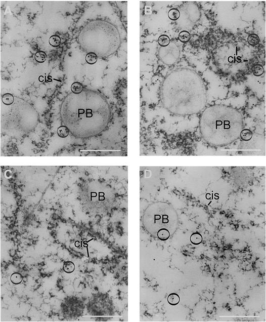
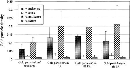
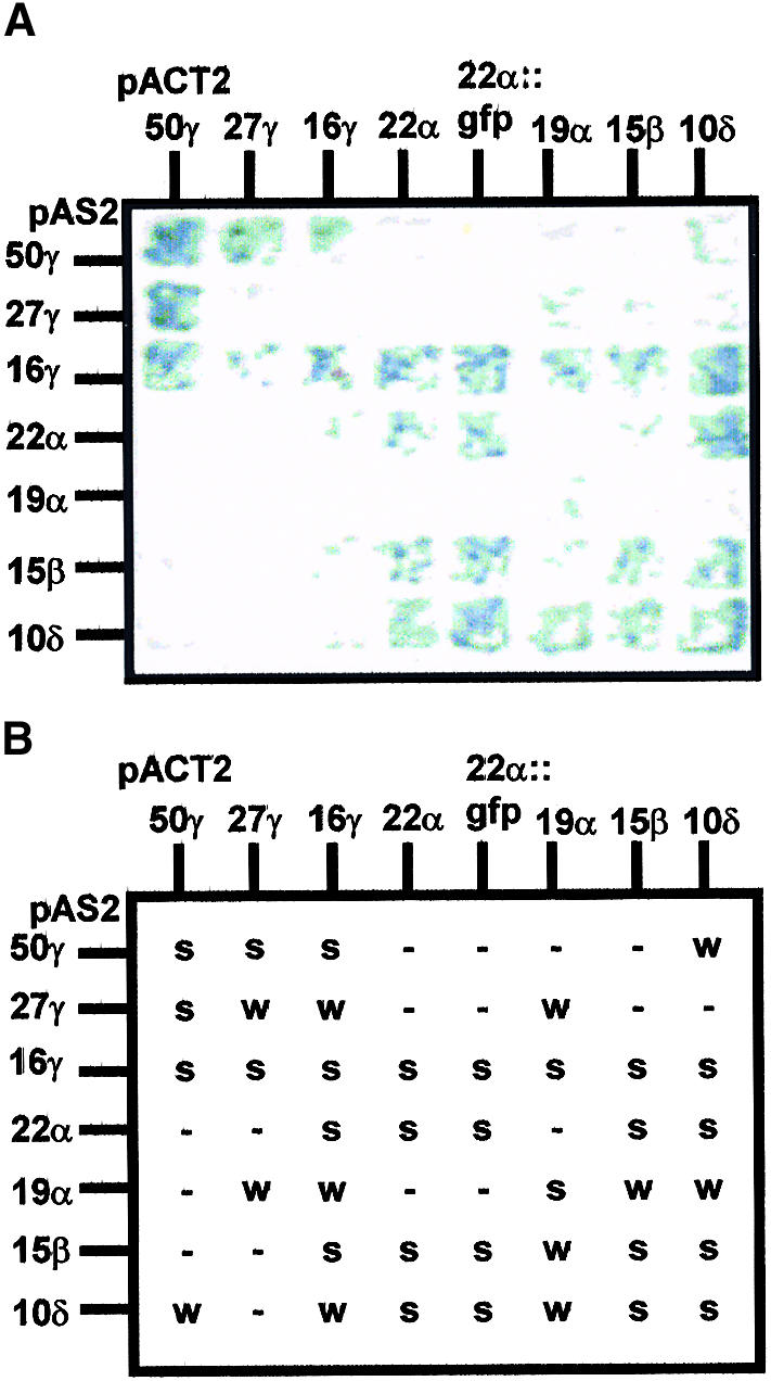
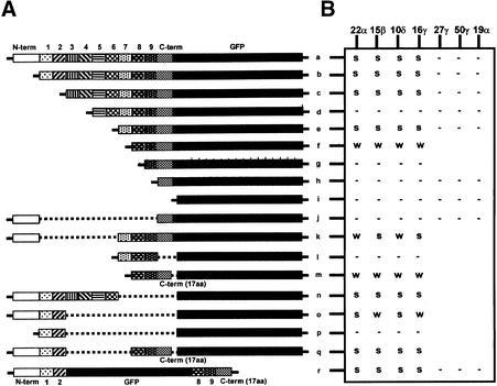
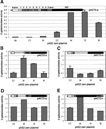
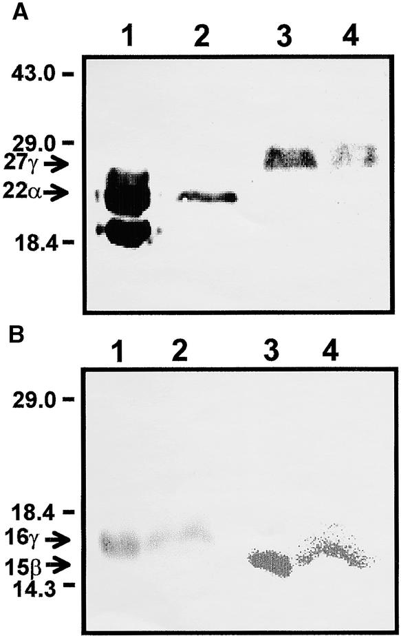
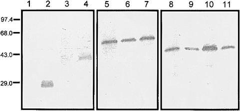
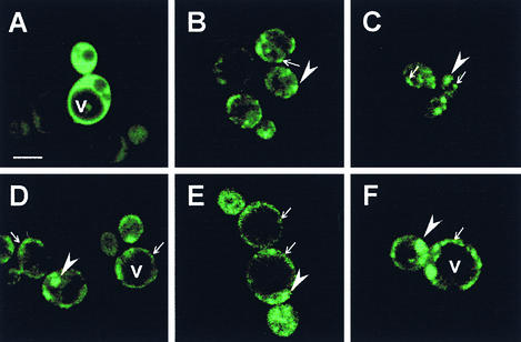
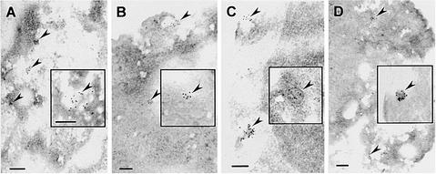
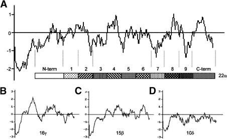
Prolamin-containing protein bodies in maize endosperm are composed of four different polypeptides, the alpha-, beta-, gamma-, and delta-zeins. The spatial organization of zeins within the protein body, as well as interactions between them, suggests that the localized synthesis of gamma-zeins could initiate and target protein body formation at specific regions of the rough endoplasmic reticulum. To investigate this possibility, we analyzed the distribution of mRNAs encoding the 22-kD alpha-zein and the 27-kD gamma-zein proteins on cisternal and protein body rough endoplasmic reticulum membranes. In situ hybridization revealed similar frequencies of the mRNAs in both regions of the endoplasmic reticulum, indicating that the transcripts are distributed more or less randomly. This finding implies that zein protein interactions determine protein body assembly. To address this question, we expressed cDNAs encoding alpha-, beta-, gamma-, and delta-zeins in the yeast two-hybrid system. We found strong interactions among the 50-, 27-, and 16-kD gamma-zeins and the 15-kD beta-zein, consistent with their colocalization in developing protein bodies. Interactions between the 19- and 22-kD alpha-zeins were relatively weak, although each of them interacted strongly with the 10-kD delta-zein. Strong interactions were detected between the alpha- and delta-zeins and the 16-kD gamma-zein and the 15-kD beta-zein; however, the 50- and 27-kD gamma-zeins did not interact with the alpha- and delta-zein proteins. We identified domains within the 22-kD alpha-zein that bound preferentially the alpha- and delta-zeins and the beta- and gamma-zeins. Affinities between zeins generally were consistent with results from immunolocalization experiments, suggesting an important role for the 16-kD gamma-zein and the 15-kD beta-zein in the binding and assembly of alpha-zeins within the protein body.


Figures

Figure 1.
Figure 1.
Localization of the 27-kD γ-Zein and the 22-kD α-Zein mRNAs in Developing Maize Endosperm. (A) Electron micrograph of an endosperm section hybridized with the 27-kD γ-zein antisense probe. (B) Electron micrograph of an endosperm section hybridized with the 22-kD α-zein antisense probe. (C) Electron micrograph of an endosperm section hybridized with the 27-kD γ-zein sense probe. (D) Electron micrograph of an endosperm section hybridized with the 22-kD α-zein sense probe. Circles are around colloidal gold particles, which appear in clusters or strings in the antisense-treated samples and usually as singlet particles in the sense-treated samples. In samples hybridized with antisense probes, label was found primarily around protein bodies (PB) or on the cisternal (cis) ER, whereas in the sense-treated samples, label was found at random locations in the cell. These observations were confirmed based on the statistical analyses in Figure 2. Bars = 1.0 μm.
Figure 2.
Figure 2.
Distribution of Colloidal Gold Particles per Micrometer of Protein Body versus Cisternal ER on Endosperm Sections Treated with 22-kD α-Zein and 27-kD γ-Zein Antisense and Sense Probes (as Illustrated in Figure 1). The method for counting gold particles is described in Methods; error bars indicate standard error. cis, cisternal; PB, protein body.
Figure 3.
Figure 3.
Assay of Zein Interactions with the Yeast Two-Hybrid System. After removal of the signal peptide sequence, zein coding regions were subcloned into the pAS2 and pACT2 plasmids of the yeast two-hybrid system and transformed reciprocally into yeast cells. (A) Yeast cells inoculated on filters and grown with X-Gal overnight. (B) Yeast grown on plates containing 3-aminotriazole for 6 days. Strong (s) indicates that colonies expressing the two indicated zeins had a positive reaction in the X-Gal filter lift assay and scored positively for growth on medium containing 3-aminotriazole compared with negative controls. Weak (w) indicates that colonies expressing the indicated zeins showed a slight color reaction in the X-Gal assay but grew slowly on selection medium containing 3-aminotriazole compared with controls. (−) indicates that no X-Gal reaction was detected.
Figure 4.
Figure 4.
Assay of Yeast Two-Hybrid Interactions between 22-kD α-Zein Deletion Mutants and Other Types of Zein Proteins. (A) Construction of the 22-kD α-zein deletion mutants in the pACT2 plasmid. White boxes correspond to the N-terminal leader following the signal peptide; dotted, hatched, and cross-hatched boxes (1 to 9) correspond to the 20–amino acid repeated peptides; the C-terminal 17 amino acids (17 aa) are indicated by solid gray boxes; and the C-terminal GFP fusion is shown by black boxes. (B) The nature of the yeast two-hybrid interaction between 22-kD α-zein deletion mutants and zein proteins. Strong (s) indicates that colonies expressing the two indicated zeins showed a positive reaction in the X-Gal filter lift assay and scored positively for growth on medium containing 3-aminotriazole compared with negative controls. Weak (w) indicates that colonies expressing the indicated zeins showed a slight color reaction in the X-Gal assay but grew slowly on selective medium containing 3-aminotriazole compared with controls. (−) indicates that no X-Gal reaction was detected.
Figure 5.
Figure 5.
Assay of β-Galactosidase Activity Resulting from Yeast Two-Hybrid Interactions between Selected 22-kD α-Zein Deletion Mutants and Other Zein Proteins. Activation of the LacZ reporter gene, measured as β-galactosidase activity with ONPG substrate, was assayed to measure the strength of the yeast two-hybrid interaction. The values shown are averages of three experiments. A diagram illustrating the structure of the 22-kD α-zein deletion mutant (see Figure 4A) in the pACT2 plasmid is provided for each assay. (A) Interaction of the complete 22-kD α-zein (construct pACT2-a) with other zeins. (B) Yeast coexpressing construct pACT2-k and the 22-kD α-zein, 16-kD γ-zein, 15-kD β-zein, and 10-kD δ-zein. (C) Yeast coexpressing construct pACT2-o and the 22-kD α-zein, 16-kD γ-zein, 15-kD β-zein, and 10-kD δ-zein. (D) Yeast coexpressing construct pACT2-q and the 22-kD α-zein, 16-kD γ-zein, 15-kD β-zein, and 10-kD δ-zein. (E) Yeast coexpressing construct pACT2-r and the 22-kD α-zein, 16-kD γ-zein, 15-kD β-zein, and 10-kD δ-zein. Error bars indicate ±sd.
Figure 6.
Figure 6.
Immunodetection of α-, β-, and γ-Zein Proteins Synthesized in Yeast. Protein extracts from yeast transformed with native zein-expressing plasmids were separated by 12% SDS-PAGE and immunoblotted with the zein antisera described below. The molecular masses of protein standards (kD) and the migration of the various zein proteins are indicated at left. (A) Immunoblot detecting the 22-kD α-zein and the 27-kD γ-zein. Lanes 1 and 3, maize endosperm extract; lane 2, yeast expressing the 22-kD α-zein; lane 4, yeast expressing the 27-kD γ-zein. (B) Immunoblot detecting the 16-kD γ-zein and the 15-kD β-zein. Lane 1 and 3, maize endosperm extract; lane 2, yeast expressing the 16-kD γ-zein; lane 4, yeast expressing the 15-kD β-zein.
Figure 7.
Figure 7.
Immunoblot Analysis of Yeast Cells Producing GFP and Zein-GFP Fusions. Yeast extracts were prepared from cells grown to an OD600 of 1.0, 17 μL of cell lysate was loaded in each lane, and the proteins were detected with a GFP monoclonal antibody. Lane 1, wild-type yeast; lane 2, yeast expressing GFP; lane 3, yeast expressing 15-kD β-zein::GFP; lane 4, yeast expressing 16-kD γ-zein::GFP; lane 5, yeast expressing 22-kD α-zein::GFP; lane 6, yeast coexpressing 22-kD α-zein::GFP and the 15-kD β-zein; lane 7, yeast coexpressing 22-kD α-zein::GFP and the 16-kD γ-zein; lane 8, yeast expressing construct pGPD-r (see Figure 8A); lanes 9, 10, and 11, yeast coexpressing construct pGPD-r and the 15-kD β-zein, 16-kD γ-zein, and 22-kD α-zein, respectably. The molecular masses (kD) of protein standards are shown at left.
Figure 8.
Figure 8.
Confocal Laser Scanning Microscopy Images of Yeast Cells Producing Maize Zein Proteins as GFP Fusions. (A) Yeast cells synthesizing GFP alone. (B) Yeast cells synthesizing 22-kD α-zein::GFP. (C) Yeast cells synthesizing 22-kD α-zein::GFP and the 15-kD β-zein. (D) Yeast cells synthesizing 22-kD α-zein::GFP and the 16-kD γ-zein. (E) Yeast cells synthesizing construct pGPD-r. (F) Yeast cells synthesizing construct pGPD-r and the 15-kD β-zein. Yeast cells expressing zein::GFP fusion proteins contained relatively small, fluorescent structures (arrows) and fluorescent clumps (arrowheads) of various shapes and sizes, whereas fluorescence was diffuse in the cytoplasm of yeast cells expressing GFP alone. The fluorescence was excluded from the large central vacuole (v). All images are at a same magnification; bar in (A) = 10 μm.
Figure 9.
Figure 9.
Immunolocalization of Zeins in Yeast Cells. (A) Yeast cells synthesizing 22-kD α-zein::GFP. (B) Yeast cells synthesizing 15-kD β-zein::GFP. (C) Yeast cells synthesizing 22-kD α-zein::GFP and the 15-kD β-zein. (D) Yeast cells synthesizing construct pGPD-r. Arrowheads indicate accretions decorated with multiple gold particles. The most prominent protein bodies found from another cell of the corresponding yeast population are shown in the insets. For the single immunolabeling shown in (A), (B), and (D), 10-nm gold particles were used. In (C), 22-kD α-zein::GFP was labeled with 10-nm gold particles, and the 15-kD β-zein was labeled with 5-nm gold particles. Bars = 200 nm.
Figure 10.
Figure 10.
Kyte-Doolittle Hydropathy Analysis of the Primary Amino Acid Sequences of Zein Proteins. (A) The 22-kD α-zein. (B) The 16-kD γ-zein. (C) The 15-kD β-zein. (D) The 10-kD δ-zein.

Similar articles

Cited by

References

    1. Abe, S., You, W., and Davies, E. (1991). Protein bodies in corn endosperm are enclosed by and enmeshed in F-actin. Protoplasma 165, 139–149.
    1. Argos, P., Pedersen, K., Marks, M.D., and Larkins, B.A. (1982). A structural model for maize zein proteins. J. Biol. Chem. 257, 9984–9990. - PubMed
    1. Bagga, S., Adams, H.P., Rodriguez, F.D., Kemp, J.D., and Sengupta-Gopalan, C. (1997). Coexpression of the maize delta-zein and beta-zein genes results in stable accumulation of delta-zein in endoplasmic reticulum-derived protein bodies formed by beta-zein. Plant Cell 9, 1683–1696. - PMC - PubMed
    1. Bai, C., and Elledge, S.J. (1996). Gene identification using the yeast two-hybrid system. Methods Enzymol. 273, 331–347. - PubMed
    1. Bar-Peled, M., Bassham, D.C., and Raikhel, N.V. (1996). Transport of proteins in eukaryotic cells: More questions ahead. Plant Mol. Biol. 32, 223–249. - PubMed

Publication types