Comparative Study
. 2002 Mar;14(3):689-702.
doi: 10.1105/tpc.010357.

The Cf-9 disease resistance protein is present in an approximately 420-kilodalton heteromultimeric membrane-associated complex at one molecule per complex

Affiliations
Comparative Study

The Cf-9 disease resistance protein is present in an approximately 420-kilodalton heteromultimeric membrane-associated complex at one molecule per complex

Susana Rivas et al. Plant Cell. 2002 Mar.

Retraction in

Abstract

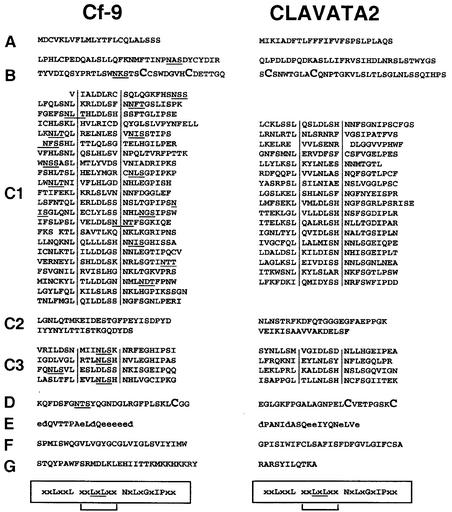
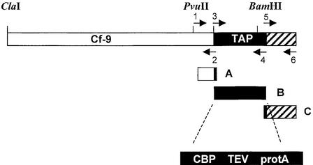
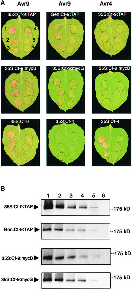
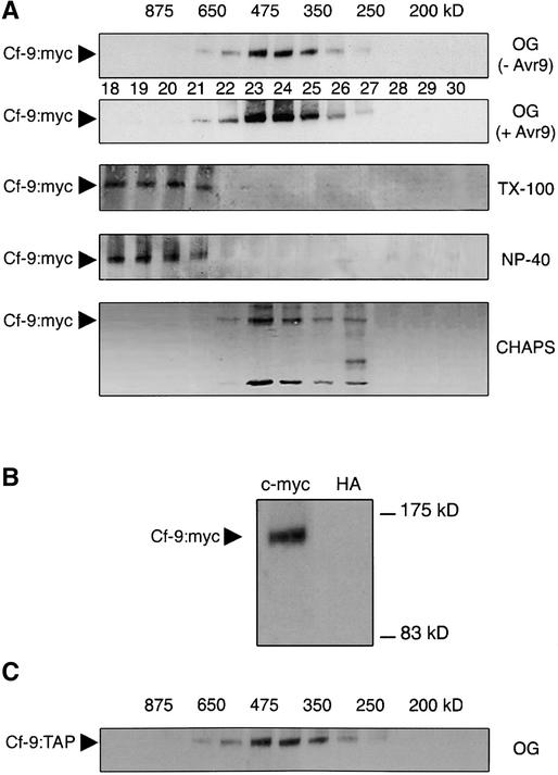
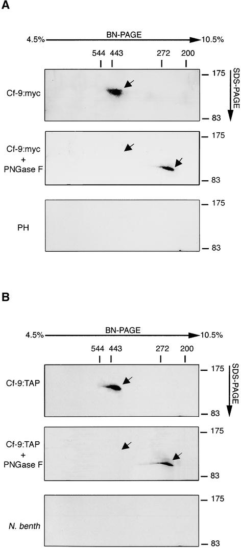
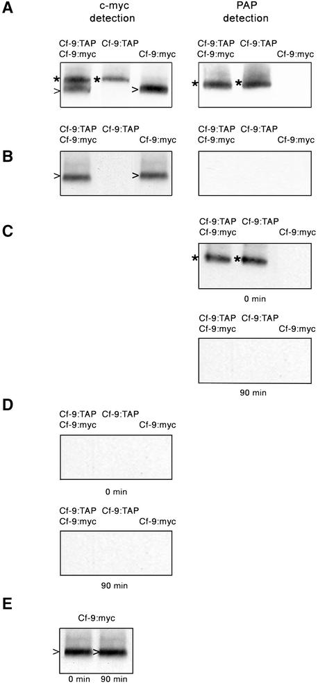
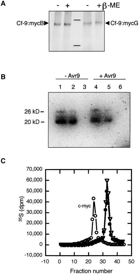
The tomato Cf-9 gene confers race-specific resistance to the fungal pathogen Cladosporium fulvum expressing the corresponding avirulence gene Avr9. In tobacco, Cf-9 confers a hypersensitive response to the Avr9 peptide. To investigate Cf-9 protein function in initiating defense signaling, we engineered a functional C-terminal fusion of the Cf-9 gene with the TAP (Tandem Affinity Purification) tag. In addition, we established a transient expression assay in Nicotiana benthamiana leaves for the production of functional Cf-9:myc and Cf-9:TAP. Transiently expressed Cf-9:myc and Cf-9:TAP proteins induced an Avr9-dependent hypersensitive response, consistent with previous results with stably transformed tobacco plants and derived cell suspension cultures expressing c-myc-tagged Cf-9. Gel filtration of microsomal fractions solubilized with octylglucoside revealed that the Cf-9 protein, either as c-myc or TAP fusions, migrated at a molecular mass of 350 to 475 kD. By using blue native gel electrophoresis, the molecular size was confirmed to be approximately 420 kD. Our results suggest that only one Cf-9 protein molecule is present in the Cf-9 complex and that Cf-9 is part of a membrane complex consisting of an additional glycoprotein partner(s). The high structural similarity between Cf proteins and Clavata2 (CLV2) of Arabidopsis, together with the similarity of molecular mass between Cf-9 and CLV complexes (420 and 450 kD, respectively), led us to investigate whether Cf-9 is integrated into membrane-associated protein complexes like those formed by CLV1 and CLV2. Unlike CLV2, the Cf-9 protein did not form disulfide-linked heterodimers, no ligand (Avr9)-dependent shift in the molecular mass of the Cf-9 complex was detected, and no Rho-GTPase-related proteins were found associated with Cf-9 under the conditions tested. Thus, Cf-9-dependent defense signaling and CLV2-dependent regulation of meristem development seem to be accomplished via distinct mechanisms, despite the structural similarity of their key components Cf-9 and CLV2.


Figures

Figure 1.
Figure 1.
Comparison of Tobacco Cf-9 and Arabidopsis CLV2 Proteins. Comparison of the amino acid sequences of Cf-9 (left) and CLV2 (right) (Jeong et al., 1999) reveals that both proteins can be subdivided into nine distinct domains (A to G) of similar structure. The amino acid sequence identity between CLV2 and Cf-9 is 21.5%. Potential NxS/T glycosylation sites are underlined in Cf-9. Domain A represents a putative signal peptide, and domain B is the mature N terminus. Cys pairs flanking the LRRs (domains C1 and C3) (Jones and Jones, 1997) are shown in larger type in domains B and D. Domains E (acidic domain with acidic residues shown in lowercase letters), F (transmembrane domain), and G (cytosolic domain) are predicted to anchor and orient Cf-9 in the cell membrane (Jones et al., 1994). The consensus sequences for plant extracellular LRRs are shown boxed and aligned below the amino acid sequences, and consensus residues with a possible β-strand configuration are underlined. Residues corresponding to the β-strand/β-turn structural motif (Kobe and Deisenhofer, 1994; Jones and Jones, 1997) are indicated by brackets and delimited by vertical lines.
Figure 2.
Figure 2.
Scheme of Cf-9:TAP. The Cf-9 sequence is represented by a white bar. The TAP tag is shown in black, with the relative positions of the calmodulin binding peptide (CBP), the TEV protease cleavage site (TEV), and the IgG binding units of protein A from S. aureus (protA) indicated within the magnified block. The 3′ untranslated sequence of Cf-9 is shown in the striped box. The TAP sequence was inserted by chimeric polymerase chain reaction at the 3′ end of the Cf-9 gene (see Methods). The three overlapping fragments used for the chimeric reaction (A, B, and C) are shown. Positions of the primers and the restriction sites used for cloning are indicated.
Figure 3.
Figure 3.
Function and Detection of Cf-9:TAP and Cf-9:myc Transiently Expressed in N. benthamiana Leaves. (A) Leaves of N. benthamiana transgenic for Avr9 (left and middle) or Avr4 (right) were infiltrated with A. tumefaciens carrying different Cf constructs, under 35S or Cf-9 endogenous promoter (Gen), at six different cell densities (1, OD600 = 0.1; 2, OD600 = 0.05; 3, OD600 = 0.02; 4, OD600 = 0.01; 5, OD600 = 0.005; 6, OD600 = 0.002), as indicated in the leaf at top left. After 5 days, the Cf-9/Avr9-dependent hypersensitive cell death reaction was observed in the Avr9 leaves infiltrated with Cf-9 constructs. (B) N. benthamiana leaves were infiltrated as in (A). Microsomal fractions were prepared 2 days after infiltration. Proteins (50 μg) were separated on a SDS-PAGE gel and analyzed by immunoblotting using a PAP or an anti-c-myc antibody for the detection of TAP- and c-myc–tagged Cf-9, respectively. Positions of c-myc Cf-9 and Cf-9:TAP are indicated by arrowheads. Numbers at top indicate the A. tumefaciens cell density used for infiltration.
Figure 4.
Figure 4.
Gel Filtration Analysis of the Cf-9:myc and Cf-9:TAP Microsomes. (A) Microsomal proteins (0.5 mg) from SLJ9161 tobacco cell suspensions were solubilized with detergents TX-100 (0.1%), Nonidet P-40 (NP-40; 0.1%), CHAPS (0.5%), or OG (40 mM; before [− Avr9] and after [+ Avr9] elicitation) for 30 min on ice, and the supernatants were subjected to gel filtration chromatography on a Sephacryl S-300 column. Two-milliliter fractions were collected, and aliquots were analyzed by SDS-PAGE and immunoblotting with an anti-myc antibody. Fraction numbers of the elution profile are indicated by the numbers between the gels. The molecular mass estimated for each fraction (in kD) is given at top. Cf-9:myc is indicated by arrowheads. (B) Immunoprecipitation of Cf-9:myc from pooled fractions of the gel filtration. Microsomes were prepared from SLJ9161 homozygous tobacco plants, solubilized with OG, and subjected to gel filtration chromatography as described in (A). Fractions containing Cf-9:myc were pooled and subjected to immunoprecipitation with a monoclonal anti-c-myc or anti-hemagglutinin (HA) antibody (control). Precipitated proteins were analyzed by SDS-PAGE and immunoblotting using a polyclonal anti-c-myc antibody. The position of Cf-9:myc is indicated by an arrowhead, and the sizes of molecular mass markers are shown at right. (C) Two N. benthamiana leaves were infiltrated with A. tumefaciens carrying 35S:Cf-9:TAP. Two days after infiltration, microsomal proteins (0.5 mg) were solubilized with 40 mM OG and subjected to gel filtration chromatography as described in (A). The molecular mass estimated for each fraction (in kD) is given at top. Cf-9:TAP is indicated by an arrowhead.
Figure 5.
Figure 5.
BN-PAGE of Solubilized Cf-9:myc and Cf-9:TAP Microsomes. (A) Microsomal proteins (0.5 mg) of SLJ9161 cell cultures (Cf-9:myc; top and middle) and Petit Havana (PH; bottom) were solubilized with 40 mM OG and 750 mM ɛ-aminocaproic acid in 50 mM bis-Tris for 30 min on ice. Aliquots of supernatants (80 μg of protein in 40 μL) were either treated with PNGase F (middle; see legend to Figure 2A) or incubated directly with 5% Coomassie blue. Proteins were separated on a 4.5 to 10.5% BN-PAGE gradient gel. Cf complexes were resolved by two-dimensional 7.5% SDS-PAGE and blotted onto nitrocellulose. Cf-9:myc protein was finally detected by immunoblot analysis of the SDS gel using an anti-c-myc antibody. The sizes of the molecular mass markers are indicated at top (BN-PAGE) and at right (SDS-PAGE). The positions of Cf-9:myc are indicated by arrows. (B) N. benthamiana (N. benth) leaves were infiltrated or not with A. tumefaciens carrying 35S:Cf-9:TAP. After 2 days, microsomal proteins (0.5 mg) of infiltrated leaves (top and middle) and non-injected leaves (bottom) were solubilized as indicated in (A). Cf-9:TAP preparations were treated or not with PNGase F (top and middle, respectively), and then BN-PAGE was conducted. Cf-9:TAP protein was detected by immunoblot analysis using the PAP antibody. The positions of Cf-9:TAP are indicated by arrows.
Figure 6.
Figure 6.
Stoichiometry of the Cf-9 Protein in the Cf-9 Complex. N. benthamiana leaves were infiltrated with A. tumefaciens expressing Cf-9:TAP and Cf-9:myc, Cf-9:TAP, or Cf-9:myc. Leaves were harvested 2 days after infiltration, and microsomal proteins were prepared. (A) Transient protein expression was analyzed by protein gel blotting with monoclonal anti-c-myc (left) and PAP antibodies (right). (B) Protein extracts were subjected to immunoprecipitation with rabbit IgG-agarose beads, immunoprecipitates were collected by centrifugation, and the non-immunoprecipitated material was analyzed again by protein gel blotting with monoclonal anti-c-myc (left) and PAP antibodies (right). (C) and (D) Immunoprecipitates were cleaved with the TEV protease, and aliquots of both IgG beads (C) and cleaved material (D) were immunoanalyzed with the PAP and the anti-c-myc antibody, respectively, before (0 min) and after (90 min) incubation with TEV enzyme. (E) A control incubation of Cf-9:myc microsomes with the TEV protease was also performed. Cf-9:myc before (0 min) and after (90 min) incubation with TEV was detected with a monoclonal anti-c-myc antibody. *, Cf-9:TAP; >, Cf-9:myc.
Figure 7.
Figure 7.
Characterization of the Cf-9:myc Complex. (A) Disulfide linkage analysis of Cf-9:mycB and Cf-9:mycG. Solubilized microsomal fractions from SLJ9171 homozygous tobacco plants (Cf-9:mycB; lanes 1 and 2) and SLJ9161 homozygous tobacco plants (Cf-9:mycG; lanes 3 and 4) were boiled in sample buffer in the presence (+) or absence (−) of β-mercaptoethanol (β-ME). Proteins were analyzed subsequently by SDS-PAGE and immunoblotting using an anti-myc antibody. Positions of Cf-9:myc are indicated by arrowheads. (B) Cf-9:myc cell cultures elicited (+) or not (−) with 15 nM synthetic Avr9 were immunoprecipitated with the anti-myc antibody. Aliquots of the incubation mixtures (lanes 1 and 4), non-immunoprecipitated proteins (lanes 2 and 5), and immunoprecipitated proteins (lanes 3 and 6) were analyzed by SDS-PAGE and blotted onto nitrocellulose. The membrane then was incubated with the GTP analog γ-35S-GTP and autoradiographed. (C) Elution profile of gel filtration analysis of Cf-9:myc and small, GTP binding proteins. Total extracts of Cf-9:myc cell cultures harvested before (closed circles) or after (open inverted triangles) elicitation with 50 ng/mL synthetic Avr9 were incubated with the GTP analog γ-35S-GTP and subjected to gel filtration. Eluted fractions were collected and analyzed for the presence of Cf-9:myc by immunoblotting (open circles) and for the incorporation of γ-35S-GTP by scintillation counting before and after elicitation (closed circles and open inverted triangles, respectively).

Similar articles

Cited by

References

    1. Arnold, I., Pfeiffer, K., Neupert, W., Stuart, R.A., and Schägger, H. (1999). ATP synthase of yeast mitochondria. J. Biol. Chem. 274, 36–40. - PubMed
    1. Bengezhal, M., Wasteneys, G., and Jones, D.A. (2000). The C-terminal dilysine motif confers endoplasmic reticulum localization to type I membrane proteins in plants. Plant Cell 12, 1179–1202. - PMC - PubMed
    1. Blatt, M.R., Grabov, A., Brearley, J., Hammond-Kosack, K., and Jones, J.D.G. (1999). K+ channels of Cf-9-transgenic tobacco guard cells as targets for Cladosporium fulvum Avr9. Plant J. 19, 453–462. - PubMed
    1. Caliebe, A., Grimm, R., Kaiser, G., Lübeck, J., Soll, J., and Heins, L. (1997). The chloroplastic protein import machinery contains a Rieske-type iron-sulfur cluster and a mononuclear iron-binding protein. EMBO J. 16, 7342–7350. - PMC - PubMed
    1. Clark, S.E., Williams, R.W., and Meyerowitz, E.M. (1997). The Clavata1 gene encodes a putative receptor kinase that controls shoot and floral meristem size in Arabidopsis. Cell 89, 575–585. - PubMed

Publication types

MeSH terms

Associated data

LinkOut - more resources