Review
. 2002 Apr;11(4):739-56.
doi: 10.1110/ps.4210102.

Natively unfolded proteins: a point where biology waits for physics

Affiliations
Review

Natively unfolded proteins: a point where biology waits for physics

Vladimir N Uversky. Protein Sci. 2002 Apr.

Abstract

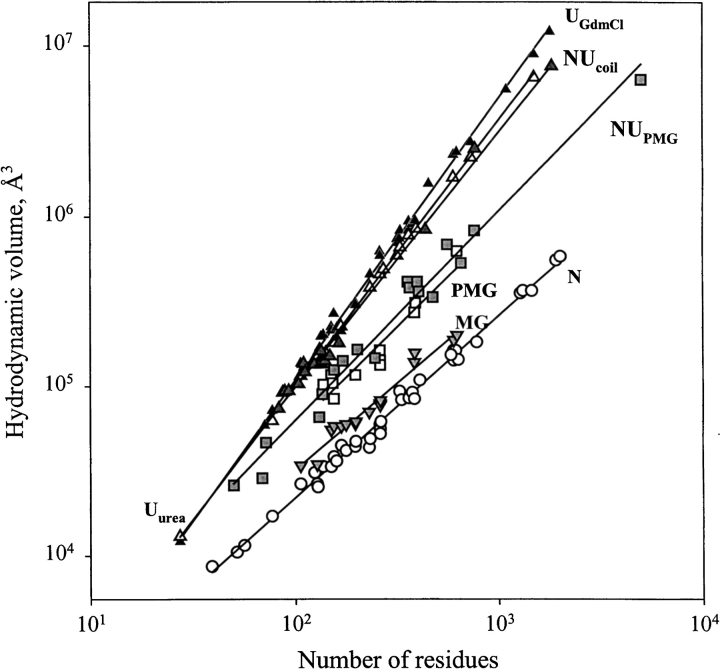
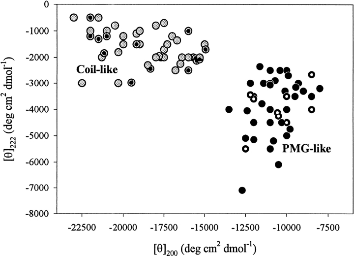
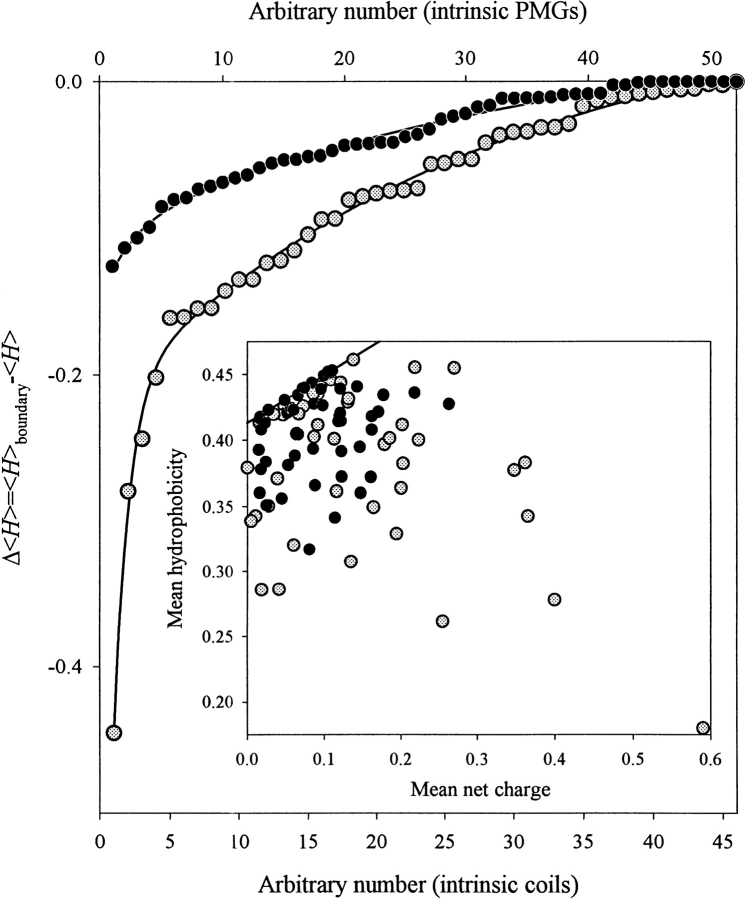
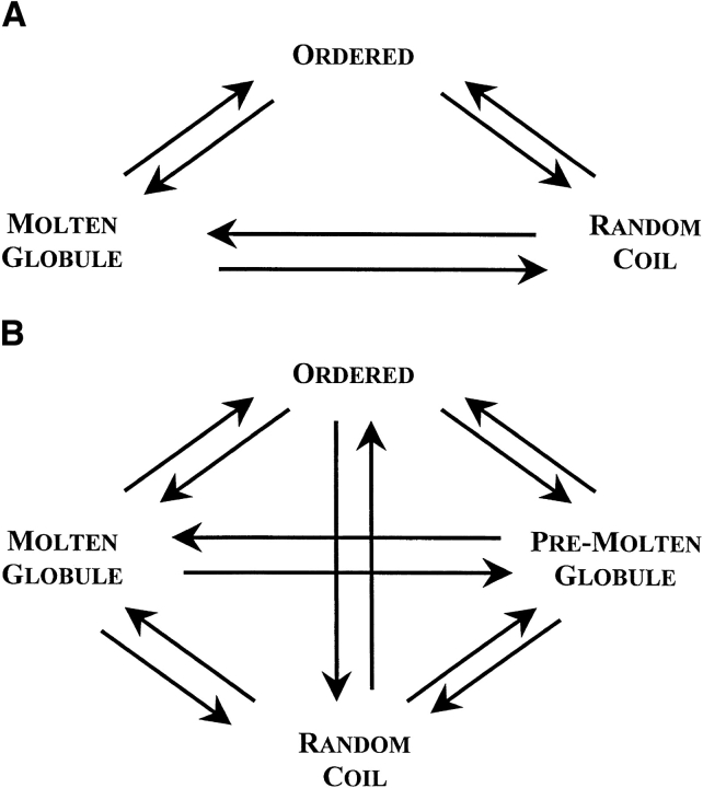
The experimental material accumulated in the literature on the conformational behavior of intrinsically unstructured (natively unfolded) proteins was analyzed. Results of this analysis showed that these proteins do not possess uniform structural properties, as expected for members of a single thermodynamic entity. Rather, these proteins may be divided into two structurally different groups: intrinsic coils, and premolten globules. Proteins from the first group have hydrodynamic dimensions typical of random coils in poor solvent and do not possess any (or almost any) ordered secondary structure. Proteins from the second group are essentially more compact, exhibiting some amount of residual secondary structure, although they are still less dense than native or molten globule proteins. An important feature of the intrinsically unstructured proteins is that they undergo disorder-order transition during or prior to their biological function. In this respect, the Protein Quartet model, with function arising from four specific conformations (ordered forms, molten globules, premolten globules, and random coils) and transitions between any two of the states, is discussed.


Figures

Fig. 1.
Fig. 1.
Protein structure–function paradigm could be considered as the big bang created universe of the modern protein science.
Fig. 2.
Fig. 2.
How the unfoldedness is encoded in protein amino acid sequence. (A) Comparison of sequences of rigid globular protein, human serum albumin (top) and intrinsically disordered protein s-antigen from Plasmodium, which was shown to be at the head of the Top 20 proteins with highest disorder scores estimated by neuronal network predictors (Romero et al. 1998b) (bottom) in terms of their composition scaled according to McCaldon and Argos (1988). (B) The unique property of natively unfolded protein sequences is a combination of low overall hydrophobicity and large net charge. Comparison of the mean hydrophobicity and the mean net charge for a set of 275 folded (black circles) and 102 natively unfolded proteins (open circles). The solid line represents the border between intrinsically unstructured and native proteins calculated using equation 1. Data for this plot are taken from Uversky et al. (2000a).
Fig. 3.
Fig. 3.
Dependencies of the hydrodynamic volume, VS, on protein polypeptide length, N, for native (open circles, N), molten globule (gray reversed triangles, MG), premolten globule (open squares, PMG), 8 M urea-unfolded (open triangles, Uurea) and 6 M GdmCl-unfolded (black triangles, UGdmCl) conformational states of globular proteins and natively unfolded proteins with coil-like (gray triangles, NUcoil) and PMG-like properties (gray squares, NUPMG). Data used to plot the dependencies for native, molten globule, premolten globule, and GdmCl-unfolded states of globular proteins are taken from Tcherkasskaya and Uversky (2001); data for natively unfolded proteins are summarized in Table 1.
Fig. 4.
Fig. 4.
Analysis of far-UV CD spectra in terms of double wavelength plot, [θ]222 versus [θ]200, allows the natively unfolded proteins division on coil-like (gray circles) and premolten globule-like subclasses (black circles). Intrinsic premolten globules and intrinsic coils for which the hydrodynamic parameters were measured (see Table 1) are marked by white-dotted and black-dotted symbols, respectively.
Fig. 5.
Fig. 5.
Comparison of the amino acid compositions of intrinsic coils (gray circles) and intrinsic premolten globules (black circles) in terms of the distance of a given sequence from the border between rigid and intrinsically unstructured proteins. The distances were calculated as Δ〈H〉 = (〈Hboundary − 〈H〉). The mean "boundary" hydrophobicity, 〈Hboundary, for a given polypeptide chain with a mean net charge 〈R〉 has been calculated using equation 1. Inset shows that proteins from both subclasses occupy the same region of the charge-hydrophobicity phase space, with intrinsic coils being more dispersed.
Fig. 6.
Fig. 6.
Extension of The Protein Trinity (A, Dunker et al. 2001) to the Protein Quartet model of protein functioning (B). In accordance with this model, function arises from four specific conformations of the polypeptide chain (ordered forms, molten globules, premolten globules, and random coils) and transitions between any of the states.

References

    1. Abercrombie, B.D., Kneale, G.G., Crane Robinson, C., Bradbury, E.M., Goodwin, G.H., Walker, J.M., and Johns, E.W. 1978. Studies on the conformational properties of the high-mobility-group chromosomal protein HMG 17 and its interaction with DNA. Eur. J. Biochem. 84 173–177. - PubMed
    1. Adler, A.J., Greenfield, N.J., and Fasman, G.D. 1973. Circular dichroism and optical rotatory dispersion of proteins and polypeptides. Methods Enzymol. 27 675–735. - PubMed
    1. Agianian, B., Leonard, K., Bonte, E., Van der Zandt, H., Becker, P.B., and Tucker, P.A. 1999. The glutamine-rich domain of the Drosophila GAGA factor is necessary for amyloid fiber formation in vitro, but not for chromatin remodelling. J. Mol. Biol. 285 527–544. - PubMed
    1. Alber, T., Gilbert, W.A., Ponzi, D.R., and Petsko, G.A. 1982. The role of mobility in the substrate binding and catalytic machinery of enzymes. Ciba Found. Symp. 93 4–24. - PubMed
    1. Alexandrescu, A.T., Abeygunawardana, C., and Shortle, D. 1994. Structure and dynamics of a denatured 131-residue fragment of staphylococcal nuclease: A heteronuclear NMR study. Biochemistry 33 1063–1072. - PubMed

LinkOut - more resources