. 2002 Apr;11(4):894-902.
doi: 10.1110/ps.4360102.

Subunit composition of a bicomponent toxin: staphylococcal leukocidin forms an octameric transmembrane pore

Affiliations

Subunit composition of a bicomponent toxin: staphylococcal leukocidin forms an octameric transmembrane pore

George Miles et al. Protein Sci. 2002 Apr.

Abstract

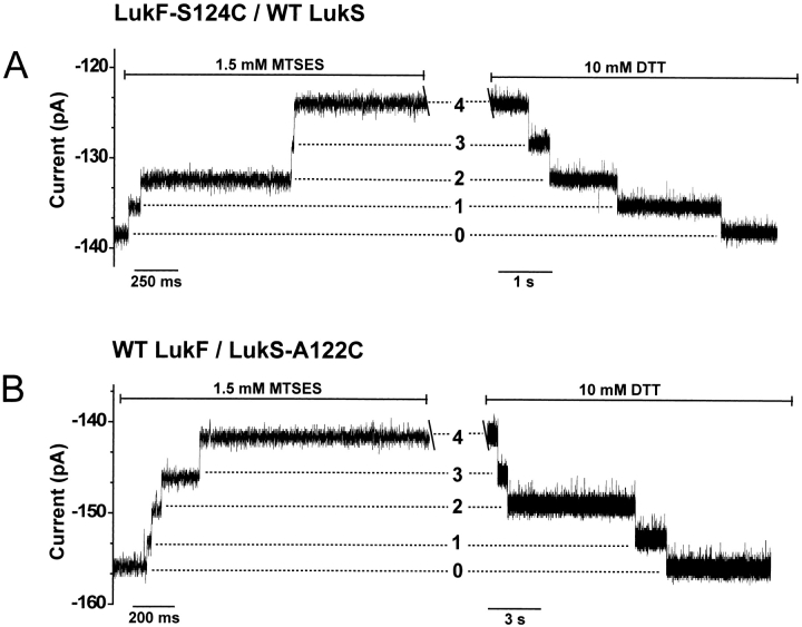
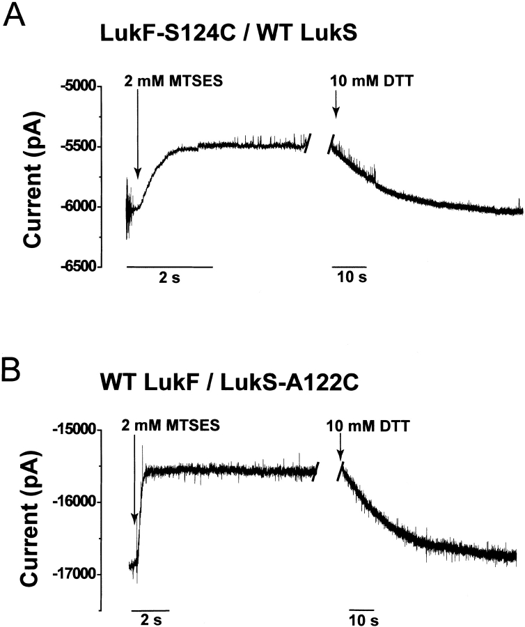
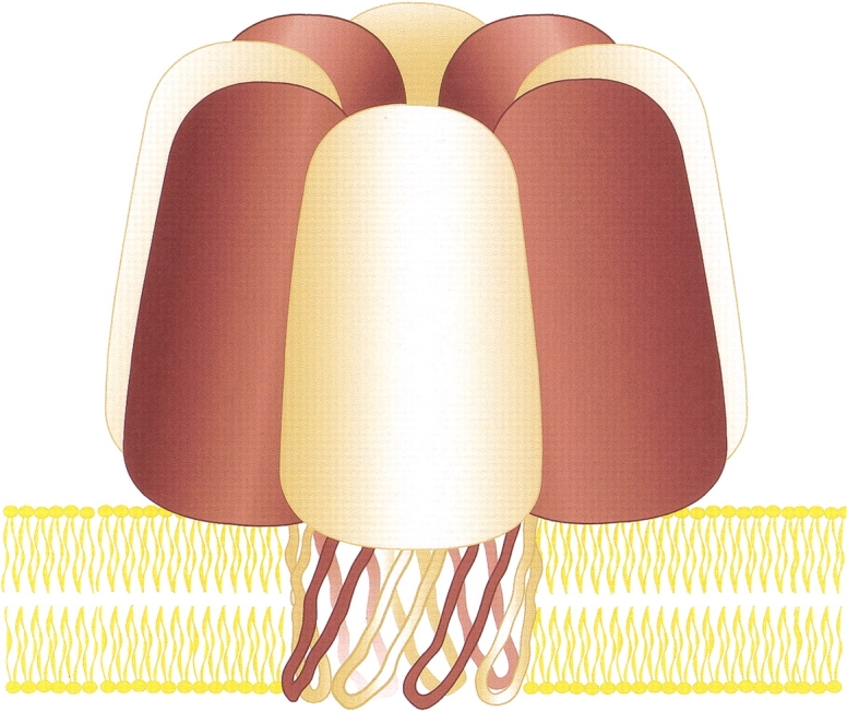
Staphylococcal leukocidin pores are formed by the obligatory interaction of two distinct polypeptides, one of class F and one of class S, making them unique in the family of beta-barrel pore-forming toxins (beta-PFTs). By contrast, other beta-PFTs form homo-oligomeric pores; for example, the staphylococcal alpha-hemolysin (alpha HL) pore is a homoheptamer. Here, we deduce the subunit composition of a leukocidin pore by two independent methods: gel shift electrophoresis and site-specific chemical modification during single-channel recording. Four LukF and four LukS subunits coassemble to form an octamer. This result in part explains properties of the leukocidin pore, such as its high conductance compared to the alpha HL pore. It is also pertinent to the mechanism of assembly of beta-PFT pores and suggests new possibilities for engineering these proteins.


Figures

Fig. 1.
Fig. 1.
Relationships between α-hemolysin (αHL) and leukocidin, members of the β-barrel pore-forming toxins (β-PFT) family. (A) Percent amino acid identity between αHL, LukF (HlgB), and LukS (HlgC). Other members of the F and S classes of leukocidin subunits are listed in the boxes. Within class F the proteins share 71%–79% identity and within class S 59%–79% identity. (B) Primary sequence alignment of αHL, LukF (HlgB), and LukS (HlgC). Identical and conserved residues are highlighted in black and gray, respectively. Residues that are identical in all three proteins are indicated with a yellow background. The residues of αHL that are located in the transmembrane β barrel, as well as the residues of LukF and LukS presumed to be in the barrel, are enclosed in the red box. The figure was generated using Clustal W 1.74 (Thompson et al. 1994). (C) Crystal structures of the LukF monomer (1LKF.pdb) and the αHL heptamer (7AHL.pdb). The secondary structure of one protomer of αHL is shown in green for comparison with LukF (maroon). The remainder of the heptamer is shown in a molecular surface representation generated by SPOCK 6.3 (Christopher 1998) and rendered using Raster3D (Merritt and Murphy 1994). (D) Possible subunit stoichiometries for the leukocidin pore. LukF and LukS are illustrated as maroon and tan spheres. The homoheptameric αHL is shown for comparison. (E) Schematic representation of the antiparallel β strands forming the β barrel of αHL and the corresponding residues of LukF and LukS. The residues are portrayed as either facing the lipid bilayer (exterior) or lining the lumen of the pore (interior). Identical residues are underscored, and identities between LukF and LukS are colored blue. Residues in red were mutated to cysteine (S124 in LukF and A122 in LukS) for modification by MTSES. The cis and trans sides of the bilayer are marked.
Fig. 1.
Fig. 1.
Relationships between α-hemolysin (αHL) and leukocidin, members of the β-barrel pore-forming toxins (β-PFT) family. (A) Percent amino acid identity between αHL, LukF (HlgB), and LukS (HlgC). Other members of the F and S classes of leukocidin subunits are listed in the boxes. Within class F the proteins share 71%–79% identity and within class S 59%–79% identity. (B) Primary sequence alignment of αHL, LukF (HlgB), and LukS (HlgC). Identical and conserved residues are highlighted in black and gray, respectively. Residues that are identical in all three proteins are indicated with a yellow background. The residues of αHL that are located in the transmembrane β barrel, as well as the residues of LukF and LukS presumed to be in the barrel, are enclosed in the red box. The figure was generated using Clustal W 1.74 (Thompson et al. 1994). (C) Crystal structures of the LukF monomer (1LKF.pdb) and the αHL heptamer (7AHL.pdb). The secondary structure of one protomer of αHL is shown in green for comparison with LukF (maroon). The remainder of the heptamer is shown in a molecular surface representation generated by SPOCK 6.3 (Christopher 1998) and rendered using Raster3D (Merritt and Murphy 1994). (D) Possible subunit stoichiometries for the leukocidin pore. LukF and LukS are illustrated as maroon and tan spheres. The homoheptameric αHL is shown for comparison. (E) Schematic representation of the antiparallel β strands forming the β barrel of αHL and the corresponding residues of LukF and LukS. The residues are portrayed as either facing the lipid bilayer (exterior) or lining the lumen of the pore (interior). Identical residues are underscored, and identities between LukF and LukS are colored blue. Residues in red were mutated to cysteine (S124 in LukF and A122 in LukS) for modification by MTSES. The cis and trans sides of the bilayer are marked.
Fig. 2.
Fig. 2.
Assembly and separation of leukocidin heteromers. (A) Separation of heteromers formed from wild-type LukS, wild-type LukF, and LukF-TL by SDS-polyacrylamide gel electrophoresis. [35S]Methionine-labeled wild type and mutant leukocidin polypeptides were synthesized in vitro by coupled transcription and translation and assembled into heteromers by including rabbit erythrocyte membranes. The concentration of LukS plasmid in the translation was equal to the total concentration of wild-type LukF and LukF-TL plasmids. The LukF and LukF-TL DNAs were mixed in the ratios indicated under the lanes. Washed membranes were solubilized in sample buffer without heating and subjected to electrophoresis in a 5% gel. An autoradiogram exposed overnight is shown. The black dots indicate the five bands that were formed. The deduced subunit compositions are listed to the right. (B) Heteromer formation from wild-type LukF and various ratios of wild-type LukS to LukS-TL, performed as described in panel A. The experiments in A and B were reproduced at least five times each. (C) Graphical representations of the leukocidin oligomers inferred from the results in B. All possible heteromeric permutations resulting from the mixtures of wild-type LukF with LukS and LukS-TL are illustrated.
Fig. 2.
Fig. 2.
Assembly and separation of leukocidin heteromers. (A) Separation of heteromers formed from wild-type LukS, wild-type LukF, and LukF-TL by SDS-polyacrylamide gel electrophoresis. [35S]Methionine-labeled wild type and mutant leukocidin polypeptides were synthesized in vitro by coupled transcription and translation and assembled into heteromers by including rabbit erythrocyte membranes. The concentration of LukS plasmid in the translation was equal to the total concentration of wild-type LukF and LukF-TL plasmids. The LukF and LukF-TL DNAs were mixed in the ratios indicated under the lanes. Washed membranes were solubilized in sample buffer without heating and subjected to electrophoresis in a 5% gel. An autoradiogram exposed overnight is shown. The black dots indicate the five bands that were formed. The deduced subunit compositions are listed to the right. (B) Heteromer formation from wild-type LukF and various ratios of wild-type LukS to LukS-TL, performed as described in panel A. The experiments in A and B were reproduced at least five times each. (C) Graphical representations of the leukocidin oligomers inferred from the results in B. All possible heteromeric permutations resulting from the mixtures of wild-type LukF with LukS and LukS-TL are illustrated.
Fig. 3.
Fig. 3.
Representative single-channel recordings of cysteine-substituted leukocidin pores reacting with MTSES. (A) LukF-S124C/wild-type LukS; (B) wild-type LukF/LukS-A122C. In both single-channel traces, there is a gap of ∼45 min between the reaction with 1.5 mM MTSES reagent and the addition of 10 mM DTT. Both reagents were added to the trans chamber. The holding potential was −60 mV and the signal was low-passed filtered at 0.5 kHz with a Bessel filter.
Fig. 4.
Fig. 4.
Typical macroscopic current recordings of leukocidin pores containing cysteine replacements during reaction with 2 mM MTSES added to the trans side of the lipid bilayer. (A) LukF-S124C/wild-type LukS; (B) wild-type LukF/LukS-A122C. In both traces, there is a gap of ∼45 min between the addition of MTSES and the reversal of the reaction with 10 mM DTT. The holding potential was −60 mV. The trans bath was stirred throughout the experiment. The signal was low-pass filtered at 1 kHz with an 8-pole Bessel filter.
Fig. 5.
Fig. 5.
Model of an octameric staphylococcal leukocidin pore. Both LukF (maroon) and LukS (tan) contribute four subunits as shown here. We suggest that the transmembrane domain of the leukocidin pore is a 16-stranded β barrel.

References

    1. Abrahams, J.P., Leslie, A.G.W., Lutter, R., and Walker, J.E. 1994. Structure at 2.8Å resolution of F1-ATPase from bovine heart mitochondria. Nature 370 621–628. - PubMed
    1. Alouf, J.E. and J.H. Freer. 1999. The comprehensive sourcebook of bacterial protein toxins, 2nd ed. Academic Press, San Diego, CA, p. 718.
    1. Archibald, J.M., Logsdon, J.M., and Doolittle, W.F. 2000. Origin and evolution of eukaryotic chaperonins: Phylogenetic evidence for ancient duplications in CCT genes. Mol. Biol. Evol. 17 1456–1466. - PubMed
    1. Baida, G., Budarina, Z.I., Kuzmin, N.P., and Solonin, A.S. 1999. Complete nucleotide sequence and molecular characterization of hemolysin II gene from Bacillus cereus. FEMS Microbiol. Lett. 180 7–14. - PubMed
    1. Bayley, H. 1997. Toxin structure: Part of a hole? Current Biol. 7 R763–R767. - PubMed

Publication types

LinkOut - more resources