

Observations on morphologic changes in the aging and degenerating human disc: secondary collagen alterations
- PMID: 11914152
- PMCID: PMC101374
- DOI: 10.1186/1471-2474-3-9
Observations on morphologic changes in the aging and degenerating human disc: secondary collagen alterations
Abstract
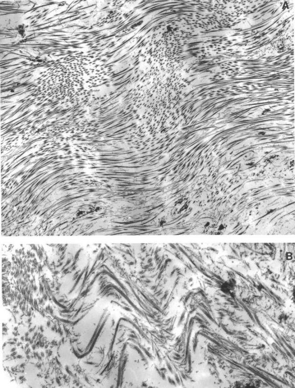
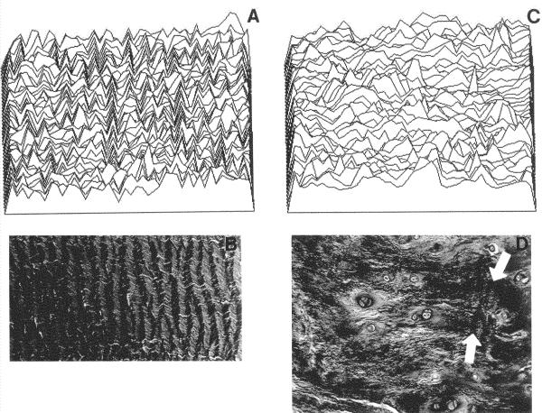
Background: In the annulus, collagen fibers that make up the lamellae have a wavy, planar crimped pattern. This crimping plays a role in disc biomechanical function by allowing collagen fibers to stretch during compression. The relationship between morphologic changes in the aging/degenerating disc and collagen crimping have not been explored.
Methods: Ultrastructural studies were performed on annulus tissue from 29 control (normal) donors (aged newborn to 79 years) and surgical specimens from 49 patients (aged 16 to 77 years). Light microscopy and specialized image analysis to visualize crimping was performed on additional control and surgical specimens. Human intervertebral disc tissue from the annulus was obtained in a prospective morphologic study of the annulus. Studies were approved by the authors' Human Subjects Institutional Review Board.
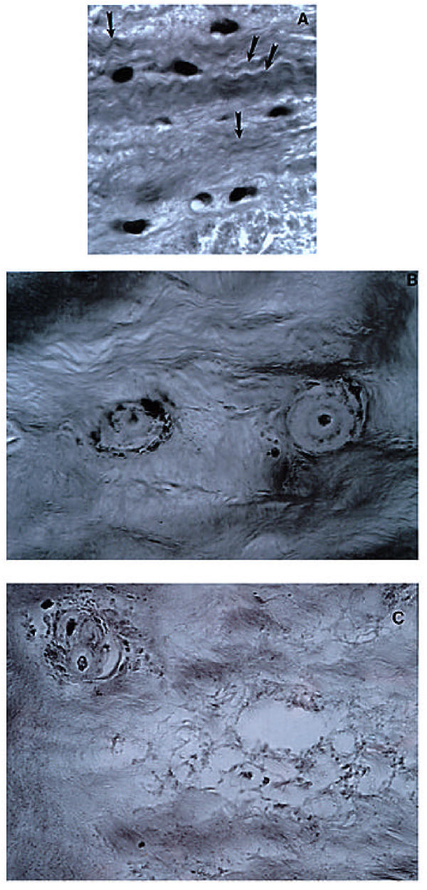
Results: Three types of morphologic changes were found to alter the crimping morphology of collagen: 1) encircling layers of unusual matrix disrupted the lamellar collagen architecture; 2) collagen fibers were reduced in amount, and 3) collagen was absent in regions with focal matrix loss.
Conclusions: Although proteoglycan loss is well recognized as playing a role in the decreased shock absorber function of the aging/degenerating disc, collagen changes have received little attention. This study suggests that important stretch responses of collagen made possible by collagen crimping may be markedly altered by morphologic changes during aging/degeneration and may contribute to the early tissue changes involved in annular tears.
Figures

References
-
- Cassidy J, Hiltner A, Baer E. Hierarchical structure of the intervertebral disc. Conn Tiss Res. 1989;23:75–88. - PubMed
-
- Cassidy J, Hiltner A, Baer E. The response of the hierarchical structure of the intervertebral disc to uniaxial compression. J Materials Science: Materials in Medicine. 1990;1:69–80.
-
- Gathercole L, Keller A. Crimp morphology in the fibre-forming collagens. Matrix. 1991;11:214–234. - PubMed
-
- Hunziker E, Ludi A, Herrmann W. Preservation of cartilage matrix proteoglycans using cationic dyes chemically related to ruthenium hexaammine trichloride. J Histochemistry Cytochemistry. 1992;40:909–917. - PubMed
LinkOut - more resources
Full Text Sources
