

Pathology of cyclodiode laser: a series of nine enucleated eyes
- PMID: 11914203
- PMCID: PMC1771068
- DOI: 10.1136/bjo.86.4.381
Pathology of cyclodiode laser: a series of nine enucleated eyes
Abstract
Aim: To study the histological effects of cyclodiode laser treatment in humans, and to compare these findings with the clinical course, treatment response, complications, and indications for enucleation.
Method: Detailed histological examination of nine enucleation specimens was undertaken in conjunction with a retrospective review of patient case notes.
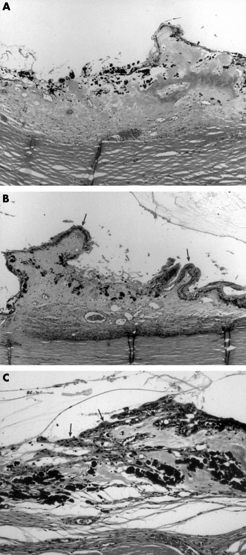
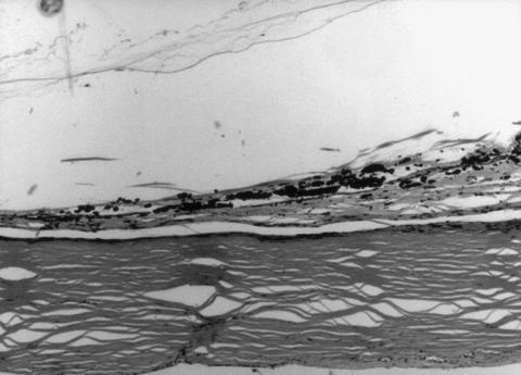
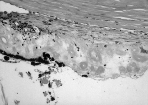
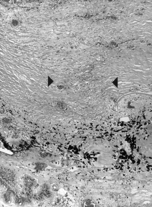
Results: Retreatments had been undertaken in three cases. Although all globes showed damage to pars plicata, intact ciliary processes within the treatment zone were present in all cases. Pars plana injury was also noted in two thirds of cases. Inflammation was mild. Ciliary epithelial proliferation was seen in most cases with increasing time following treatment, in a disorganised pattern, without replication of the ciliary epithelial bilayer. No regeneration of the ciliary processes with fibrovascular cores was found. The three patients with good IOP control at enucleation had all had multiple diode treatments. Neither phthisis nor sympathetic ophthalmia was seen.
Conclusions: Diode laser cyclophotocoagulation produces very characteristic injury to pars plicata, which frequently extends into pars plana, but with only mild persisting inflammation. Ciliary processes are, however, frequently spared within the treatment zone and may account for early or late treatment failure.
Figures
References
-
- Brindley G, Shields MB. Value and limitation of cyclocryotherapy. Graefes Arch Clin Exp Ophthalmol 1986;224:545–8. - PubMed
-
- Wagle NS, Freedman SF, Buckley EG, et al. Long-term outcome of cyclocryotherapy for refractory pediatric glaucoma. Ophthalmology 1998;105:1921–6. - PubMed
-
- Sabates R. Choroiditis compatible with the histopathologic diagnosis of sympathetic ophthalmia following cyclocryotherapy of neovascular glaucoma. Ophthalmic Surg 1988;19:176–82. - PubMed
-
- Harrison TJ. Sympathetic ophthalmia after cyclocryotherapy of neovascular glaucoma without ocular penetration. Ophthalmic Surg 1993;24:44–6. - PubMed
-
- Biswas J, Fogla R. Sympathetic ophthalmia following cyclocryotherapy with histopathologic correlation. Ophthalmic Surg Lasers 1996;27:1035–8. - PubMed
MeSH terms
LinkOut - more resources
Full Text Sources
Medical
Miscellaneous