Systemic inflammation and innate immune response in patients with previous anterior uveitis
- PMID: 11914210
- PMCID: PMC1771091
- DOI: 10.1136/bjo.86.4.412
Systemic inflammation and innate immune response in patients with previous anterior uveitis
Abstract
Aim: To determine the presence of systemic inflammation and innate immune responsiveness of patients with a history of acute anterior uveitis but no signs of ocular inflammation at the time of recruitment.
Methods: Tumour necrosis factor alpha (TNF-alpha) production in response to bacterial lipopolysaccharide (LPS) was studied using whole blood culture assay; levels of TNF-alpha in culture supernatants, and soluble interleukin 2 receptor (sIL-2R) in serum were determined by chemiluminescent immunoassay (Immulite); monocyte surface expression of CD11b, CD14, and CD16 and the proportion of monocyte subsets CD14(bright)CD16(-) and CD14(dim)CD16(+) were studied with three colour whole blood flow cytometry; and serum C reactive protein (CRP) levels were determined using immunonephelometric high sensitivity CRP assay.
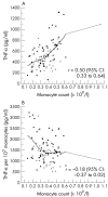
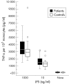
Results: The CRP level (median, interquartile range) was significantly higher in 56 patients with previous uveitis than in 37 controls (1.59 (0.63 to 3.47) microg/ml v 0.81 (0.32 to 2.09) microg/ml; p=0.008). The TNF-alpha concentration of the culture media per 10(5) monocytes was significantly higher in the patient group than in the control group in the presence of LPS 10 ng/ml (1473 (1193 to 2024) pg/ml v 1320 (935 to 1555) pg/ml; p=0.012) and LPS 1000 ng/ml (3280 (2709 to 4418) pg/ml v 2910 (2313 to 3358) pg/ml; p=0.011). The background TNF-alpha release into the culture media was low in both groups. CD14 expression of CD14(bright)CD16(-) monocytes, defined as antibody binding capacity (ABC), was similar for the patients and controls (22,839 (21,038 to 26,020) ABC v 21,657 (19,854 to 25,646) ABC).
Conclusions: Patients with previous acute anterior uveitis show high innate immune responsiveness that may play a part in the development of ocular inflammation.
Figures


References
-
- Bloch-Michel E, Nussenblatt RB. International uveitis study group recommendations for the evaluation of intraocular inflammatory disease. Am J Ophthalmol 1987;103:234–5. - PubMed
-
- Rosenbaum JT. Acute anterior uveitis and spondyloarthropathies. Rheum Dis Clin North Am 1992;18:143–51. - PubMed
-
- Rosenbaum JT, McDevitt HO, Guss RB, et al. Endotoxin-induced uveitis in rats as a model for human disease. Nature 1980;268:611–13. - PubMed
-
- Scheinberg MA, Ikejiri M, Silva MH, et al. Interleukin-2 receptor membrane in bound and soluble form in the aqueous humor and peripheral blood of patients with acute untreated uveitis. J Rheumatol 1992;19:1362–3. - PubMed
-
- Martin CM, Lacomba MS, Molina CIS, et al. Levels of soluble ICAM-1 and soluble IL-2R in the serum and aqueous humor of uveitis patients. Curr Eye Res 2000;4:287–92. - PubMed
Publication types
MeSH terms
Substances
LinkOut - more resources
Full Text Sources
Research Materials
Miscellaneous
