

Limited macular translocation with scleral retraction suture
- PMID: 11914214
- PMCID: PMC1771107
- DOI: 10.1136/bjo.86.4.434
Limited macular translocation with scleral retraction suture
Abstract
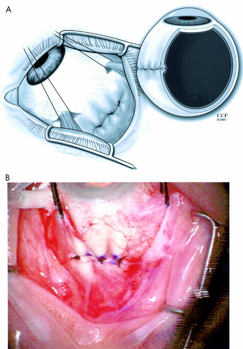
Background/aims: Macular translocation with scleral imbrication is a new technique for treating subfoveal choroidal neovascular membranes (CNV). This procedure shortens the sclera but may result in a minimal decrease in the internal circumference of the globe and limits the amount of foveal displacement. The authors propose a new scleral retraction suture aimed at decreasing the internal circumference of the globe in an effort to increase foveal displacement.
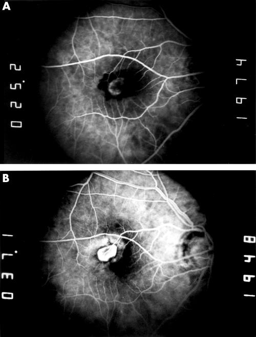
Methods: Using a cadaver model, they compared the amount of scleral shortening using a standard scleral imbrication technique and a modified three suture scleral retraction technique. Sections of the globes were digitised and specialised software was used to estimate the amount of scleral shortening. Three patients with subfoveal choroidal neovascularisation underwent limited macular translocation using pars plana vitrectomy and macular detachment with the modified scleral suture technique. The main outcome measures were visual acuity, foveal displacement, and complications.
Results: In the cadaver model, the scleral retraction suture resulted in a flatter internal scleral fold compared to the standard suture technique and created approximately 890 microm of effective scleral shortening. In the patients who underwent macular translocation and laser photocoagulation of the CNV, visual acuity improved in two patients and worsened in one patient. The range of foveal displacement was 1400-2400 microm.
Conclusion: The foveal displacements achieved in this limited study compared to median displacement previously published using standard suture techniques demonstrates that the scleral retraction suture technique may be a useful adjunct to limited macular translocation. The advantage of this type of suture in conjunction with translocation may depend on the effective scleral shortening offered by this retraction suture.
Figures


References
-
- Ferris FL III, Fine SL, Hyman LA. Age-related macular degeneration and blindness due to neovascular maculopathy. Arch Ophthalmol 1984;102:1640–2. - PubMed
-
- Leibowitz H, Krueger D, Maunder L, et al. The Framingham eye study monograph: an ophthalmological study of cataract, glaucoma, diabetic retinopathy, macular degeneration, and visual acuity in a general population of 2631 adults. Surv Ophthalmol 1973–5;24:335–610. - PubMed
-
- Macular Photocoagulation Study Group. Laser photocoagulation of subfoveal neovascular lesions in age-related macular degeneration. Arch Ophthalmol 1993;111:1200–9. - PubMed
-
- Macular Photocoagulation Study Group. Visual outcome after laser photocoagulation for subfoveal choroidal neovascularization secondary to age-related macular degeneration. Arch Ophthalmol 1994;112:480–8. - PubMed
-
- Treatment of Age-Related Macular Degeneration with Photodynamic Therapy Study Group. Photodynamic therapy of subfoveal choroidal neovascularization in age-related macular degeneration with verteporfin. Arch Ophthalmol 1999;117:1329–45. - PubMed
Publication types
MeSH terms
LinkOut - more resources
Full Text Sources
Medical
Miscellaneous