. 2002 Mar 15;16(6):693-706.
doi: 10.1101/gad.952602.

Isolation, immortalization, and characterization of a human breast epithelial cell line with stem cell properties

Affiliations

Isolation, immortalization, and characterization of a human breast epithelial cell line with stem cell properties

Thorarinn Gudjonsson et al. Genes Dev. .

Abstract

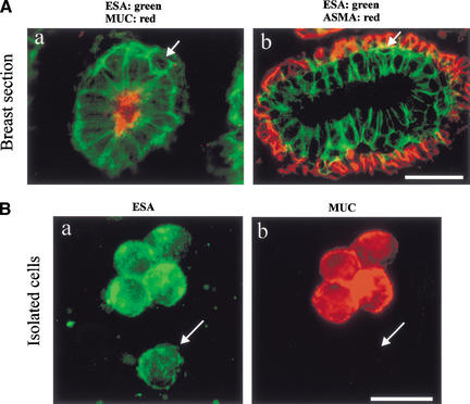
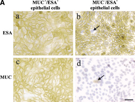
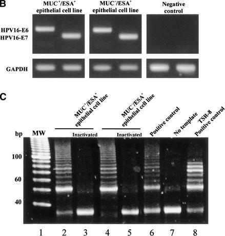
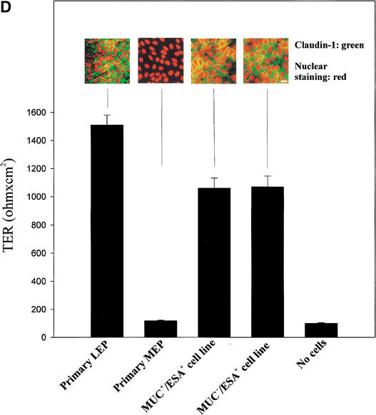
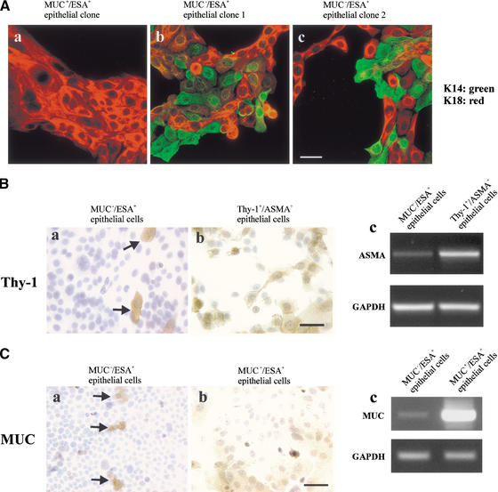
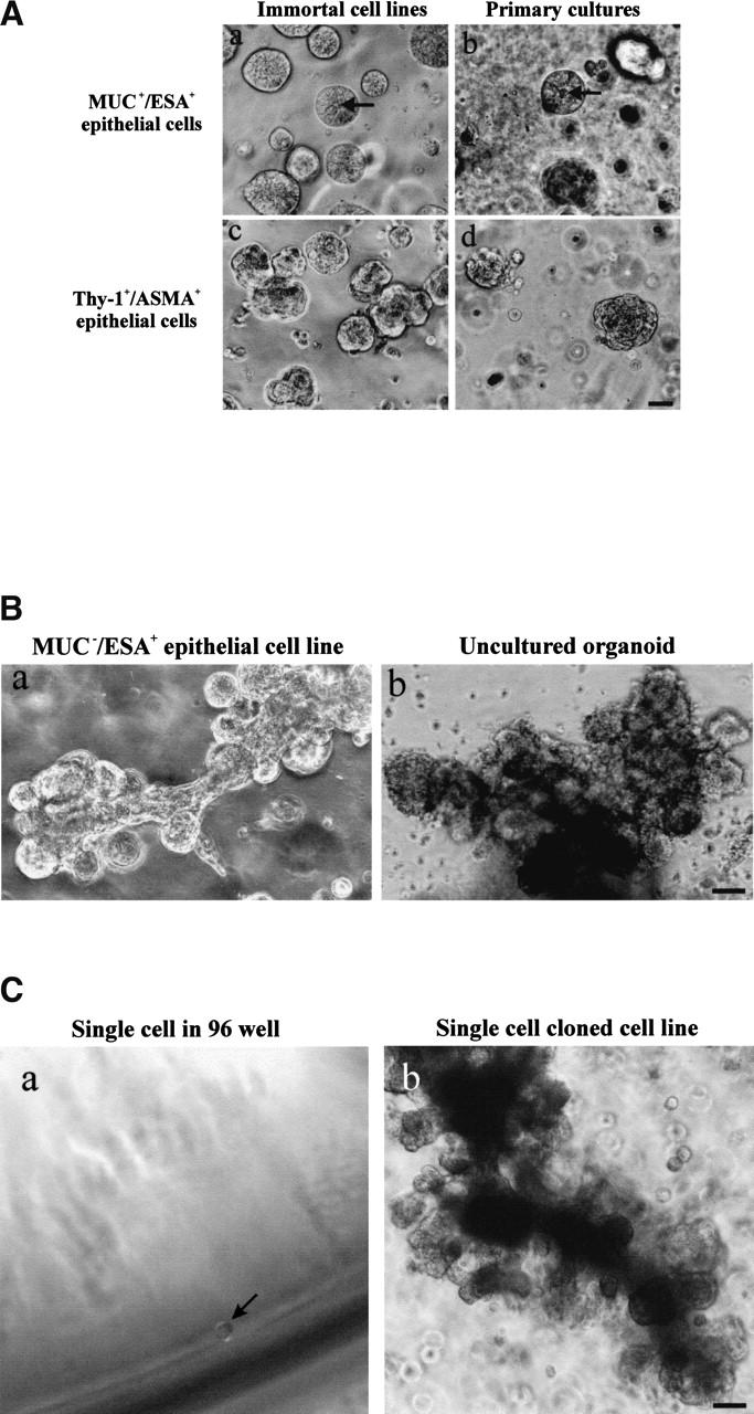
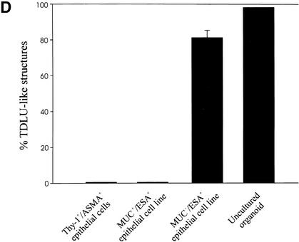
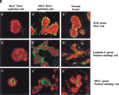
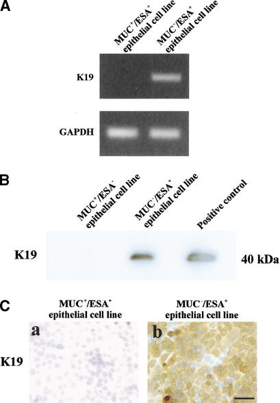
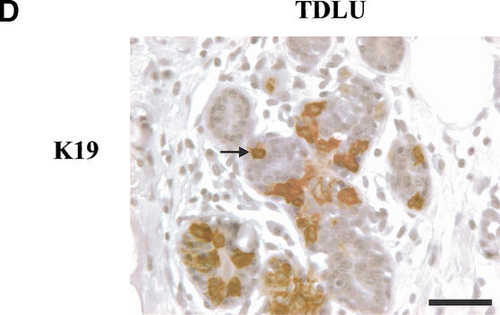
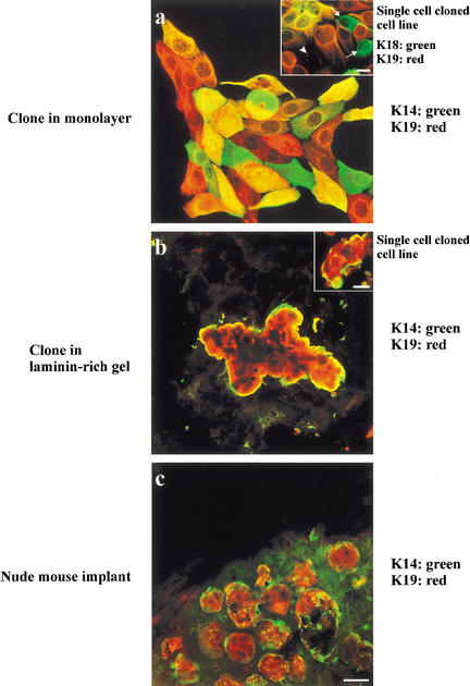
The epithelial compartment of the human breast comprises two distinct lineages: the luminal epithelial and the myoepithelial lineage. We have shown previously that a subset of the luminal epithelial cells could convert to myoepithelial cells in culture signifying the possible existence of a progenitor cell. We therefore set out to identify and isolate the putative precursor in the luminal epithelial compartment. Using cell surface markers and immunomagnetic sorting, we isolated two luminal epithelial cell populations from primary cultures of reduction mammoplasties. The major population coexpresses sialomucin (MUC(+)) and epithelial-specific antigen (ESA(+)) whereas the minor population has a suprabasal position and expresses epithelial specific antigen but no sialomucin (MUC(-)/ESA(+)). Two cell lines were further established by transduction of the E6/E7 genes from human papilloma virus type 16. Both cell lines maintained a luminal epithelial phenotype as evidenced by expression of the tight junction proteins, claudin-1 and occludin, and by generation of a high transepithelial electrical resistance on semipermeable filters. Whereas in clonal cultures, the MUC(+)/ESA(+) epithelial cell line was luminal epithelial restricted in its differentiation repertoire, the suprabasal-derived MUC(-)/ESA(+) epithelial cell line was able to generate itself as well as MUC(+)/ESA(+) epithelial cells and Thy-1(+)/alpha-smooth muscle actin(+) (ASMA(+)) myoepithelial cells. The MUC(-)/ESA(+) epithelial cell line further differed from the MUC(+)/ESA(+) epithelial cell line by the expression of keratin K19, a feature of a subpopulation of epithelial cells in terminal duct lobular units in vivo. Within a reconstituted basement membrane, the MUC(+)/ESA(+) epithelial cell line formed acinus-like spheres. In contrast, the MUC(-)/ESA(+) epithelial cell line formed elaborate branching structures resembling uncultured terminal duct lobular units both by morphology and marker expression. Similar structures were obtained by inoculating the extracellular matrix-embedded cells subcutaneously in nude mice. Thus, MUC(-)/ESA(+) epithelial cells within the luminal epithelial lineage may function as precursor cells of terminal duct lobular units in the human breast.


Figures

Figure 1
Figure 1
Identification of MUC/ESA+ luminal epithelial cells in the breast. (A) MUC/ESA+ epithelial cells belong to the luminal epithelial lineage. (a) Double-labeling immunofluorescence staining of epithelial specific antigen (ESA) and sialomucin (MUC1). The arrow indicates an example of an epithelial cell that does not appear to reach the lumen and fails to express sialomucin. (b) Double-labeling immunofluorescence staining of ESA and α-smooth muscle actin (ASMA). Note that the suprabasal epithelial cells (arrow) are resting on a layer of ASMA+ myoepithelial cells. Bar, 20 μm. (B) A subset of cells within the luminal epithelial lineage is sialomucin-negative. Uncultured, trypsinized breast epithelial cells were double-stained to demonstrate ESA (green) and sialomucin (red). Whereas the majority of cells were MUC+/ESA+, a small fraction was MUC/ESA+ (arrow). Bar, 20 μm.
Figure 2
Figure 2
Isolation, immortalization and characterization of MUC+/ESA+ and MUC/ESA+ epithelial cells. (A) The MUC+/ESA+ and MUC/ESA+ epithelial cells differ by sialomucin expression. Immunoperoxidase staining with ESA (a,b) and MUC1 (c,d) of the MUC+/ESA+ (a,c) and the MUC/ESA+ epithelial cell line (b,d). Note that whereas the MUC+/ESA+ epithelial cell line is homogenous in its staining pattern and expressed sialomucin (MUC1), the MUC/ESA+ epithelial cell line is heterogeneous (arrows) and essentially negative for sialomucin. Bar, 50 μm. (B) The cell lines express E6 and E7 stably. RT–PCR of HPV16 E6 and E7 show that both the MUC+/ESA+ and the MUC/ESA+ epithelial cells are stably transduced. (C) The cell lines exhibit telomerase activity. TRAP assay of equal numbers of MUC+/ESA+ and MUC/ESA+ epithelial cell lines showed telomerase activity in transduced cells. (Lane 1) Molecular weight markers; (lane 2) MUC+/ESA+ epithelial cell line; (lane 4) MUC/ESA+ epithelial cell line; (lanes 3 and 5) heat inactivated negative control of the cell lines; (lane 6) positive control pellet; (lane 7) negative control without cell lysate; (lane 8) positive control TSR8. (D) Both the MUC+/ESA+ and MUC/ESA+ epithelial cell lines belong to the luminal epithelial lineage. Confluent cultures were plated on Transwell filters and assayed for transepithelial resistance (TER), and parallel monolayer cultures were double-stained for claudin-1 as well as propidium iodide to visualize the nuclei (insets). Primary luminal epithelial cells (LEP) and myoepithelial cells (MEP) are readily discriminated by TER and claudin-1 expression. Bar, 20 μm.
Figure 2
Figure 2
Isolation, immortalization and characterization of MUC+/ESA+ and MUC/ESA+ epithelial cells. (A) The MUC+/ESA+ and MUC/ESA+ epithelial cells differ by sialomucin expression. Immunoperoxidase staining with ESA (a,b) and MUC1 (c,d) of the MUC+/ESA+ (a,c) and the MUC/ESA+ epithelial cell line (b,d). Note that whereas the MUC+/ESA+ epithelial cell line is homogenous in its staining pattern and expressed sialomucin (MUC1), the MUC/ESA+ epithelial cell line is heterogeneous (arrows) and essentially negative for sialomucin. Bar, 50 μm. (B) The cell lines express E6 and E7 stably. RT–PCR of HPV16 E6 and E7 show that both the MUC+/ESA+ and the MUC/ESA+ epithelial cells are stably transduced. (C) The cell lines exhibit telomerase activity. TRAP assay of equal numbers of MUC+/ESA+ and MUC/ESA+ epithelial cell lines showed telomerase activity in transduced cells. (Lane 1) Molecular weight markers; (lane 2) MUC+/ESA+ epithelial cell line; (lane 4) MUC/ESA+ epithelial cell line; (lanes 3 and 5) heat inactivated negative control of the cell lines; (lane 6) positive control pellet; (lane 7) negative control without cell lysate; (lane 8) positive control TSR8. (D) Both the MUC+/ESA+ and MUC/ESA+ epithelial cell lines belong to the luminal epithelial lineage. Confluent cultures were plated on Transwell filters and assayed for transepithelial resistance (TER), and parallel monolayer cultures were double-stained for claudin-1 as well as propidium iodide to visualize the nuclei (insets). Primary luminal epithelial cells (LEP) and myoepithelial cells (MEP) are readily discriminated by TER and claudin-1 expression. Bar, 20 μm.
Figure 3
Figure 3
Evidence for multipotency in the MUC/ESA+ epithelial cell line. (A) Double-staining with luminal epithelial K18 and myoepithelial K14 in clones of the MUC+/ESA+ epithelial cell line and the MUC/ESA+ epithelial cell line. Clonal cultures of the MUC+/ESA+ (a) and the MUC/ESA+ epithelial cells (b,c) were double-stained with keratin K18 and K14. No evidence for myoepithelial cells was found in any of the MUC+/ESA+ clones. Conversely, a mixture of cells was always present in the MUC/ESA+ epithelial clones. Bar, 40 μm. (B) Evidence of spontaneous maturation to Thy-1+/ASMA+ myoepithelial cells. Immunoperoxidase staining of Thy-1, a marker for myoepithelial cells, in cultures of MUC/ESA+ epithelial cells before (a) and after (b) purification in a Thy-1 retaining column. The spontaneous occurrence of Thy-1 stained cells is limited to <1% (arrows). However, on purification, a myoepithelial subline can be obtained that also expresses ASMA (c). Bar, 50 μm. (C) Evidence for maturation to MUC+/ESA+ epithelial cells. MUC/ESA+ epithelial cells were cleared of sialomucin-positive cells and stained for sialomucin after 2 wk (a, arrows), and after further sorting of the newly formed sialomucin-positive cells (b). The MUC stainings was confirmed by RT–PCR (c). Bar, 50 μm.
Figure 4
Figure 4
MUC/ESA+ epithelial cells give rise to terminal duct lobular units (TDLU). (A) MUC+/ESA+ epithelial- and Thy-1+/ASMA+ myoepithelial cells make colonies with distinct morphologies in a laminin-rich gel. Immortalized (a,c) and primary (b,d) MUC+/ESA+ epithelial cells (a,b) and Thy-1+/ASMA+ myoepithelial cells (c,d) were embedded as single cells in a laminin rich gel. Both MUC+/ESA+ epithelial and Thy-1+/ASMA+ myoepithelial cell lines resembled the corresponding primary cells. Whereas the luminal epithelial cells formed acinus-like spheres with a central lumen, the myoepithelial cells formed irregular solid clusters of cells. Bar, 25 μm. (B) MUC/ESA+ epithelial cells make an elaborate TDLU-like structure in a laminin-rich gel. MUC/ESA+ epithelial cells (a) were embedded as single cells in a laminin-rich gel and compared with the morphology of freshly isolated, uncultured TDLU organoids (b). Both consist of small branching ductules terminating in globular acinus-like structures. Bar, 50 μm. (C) Single cell cloned MUC/ESA+ cells maintain the ability to make TDLU-like structures in a laminin-rich gel. Single cell cloning was verified by the demonstration of only one cell in a 96 well using a 10× objective (Ca; arrow). Single cell clone ‘TH123‘ embedded as single cells in Matrigel could give rise to TDLU-like structures by 6 d (b). Bar, 50 μm. (D) Quantification of TDLU-like structures in laminin-rich gels. MUC+/ESA+ and MUC/ESA+ epithelial cells and Thy-1+/ASMA+ myoepithelial cells were embedded inside laminin-rich gels and allowed to grow for 12 d and compared to uncultured organoids. The number of TDLU-like structures was quantified by phase contrast microscopy. (E) MUC/ESA+ epithelial colonies in laminin-rich gels resemble TDLU in vivo. Sections of laminin-rich gels containing MUC+/ESA+ (left column) and MUC/ESA+ epithelial cells (middle column) were compared with sections of normal breast tissue (right column) and double-stained for ESA and keratin K14 (a–a"), propidium iodide and laminin-1 (b–b") and propidium iodide and sialomucin (c–c"). Only the MUC/ESA+ epithelial cells showed a differentiation pattern reminiscent of normal breast tissue. Bar, 15 μm.
Figure 4
Figure 4
MUC/ESA+ epithelial cells give rise to terminal duct lobular units (TDLU). (A) MUC+/ESA+ epithelial- and Thy-1+/ASMA+ myoepithelial cells make colonies with distinct morphologies in a laminin-rich gel. Immortalized (a,c) and primary (b,d) MUC+/ESA+ epithelial cells (a,b) and Thy-1+/ASMA+ myoepithelial cells (c,d) were embedded as single cells in a laminin rich gel. Both MUC+/ESA+ epithelial and Thy-1+/ASMA+ myoepithelial cell lines resembled the corresponding primary cells. Whereas the luminal epithelial cells formed acinus-like spheres with a central lumen, the myoepithelial cells formed irregular solid clusters of cells. Bar, 25 μm. (B) MUC/ESA+ epithelial cells make an elaborate TDLU-like structure in a laminin-rich gel. MUC/ESA+ epithelial cells (a) were embedded as single cells in a laminin-rich gel and compared with the morphology of freshly isolated, uncultured TDLU organoids (b). Both consist of small branching ductules terminating in globular acinus-like structures. Bar, 50 μm. (C) Single cell cloned MUC/ESA+ cells maintain the ability to make TDLU-like structures in a laminin-rich gel. Single cell cloning was verified by the demonstration of only one cell in a 96 well using a 10× objective (Ca; arrow). Single cell clone ‘TH123‘ embedded as single cells in Matrigel could give rise to TDLU-like structures by 6 d (b). Bar, 50 μm. (D) Quantification of TDLU-like structures in laminin-rich gels. MUC+/ESA+ and MUC/ESA+ epithelial cells and Thy-1+/ASMA+ myoepithelial cells were embedded inside laminin-rich gels and allowed to grow for 12 d and compared to uncultured organoids. The number of TDLU-like structures was quantified by phase contrast microscopy. (E) MUC/ESA+ epithelial colonies in laminin-rich gels resemble TDLU in vivo. Sections of laminin-rich gels containing MUC+/ESA+ (left column) and MUC/ESA+ epithelial cells (middle column) were compared with sections of normal breast tissue (right column) and double-stained for ESA and keratin K14 (a–a"), propidium iodide and laminin-1 (b–b") and propidium iodide and sialomucin (c–c"). Only the MUC/ESA+ epithelial cells showed a differentiation pattern reminiscent of normal breast tissue. Bar, 15 μm.
Figure 5
Figure 5
The MUC/ESA+ cells are keratin K19-positive similar to a subpopulation of cells in TDLU in vivo. (A) MUC+/ESA+ epithelial- and MUC/ESA+ epithelial cells differ by expression of mRNA for keratin K19. RT–PCR of keratin K19 in MUC+/ESA+ epithelial- and MUC/ESA+ epithelial cells. (B) MUC+/ESA+ epithelial- and MUC/ESA+ epithelial cells differ by expression of keratin K19. Immunoblot of keratin K19 of protein lysates from MUC+/ESA+ epithelial- and MUC/ESA+ epithelial cells. (C) Keratin K19 staining in cultures of MUC+/ESA+- and MUC/ESA+-epithelial cells. Cultures were stained for keratin K19 by immunoperoxidase and counterstained with hematoxylin. Whereas the MUC+/ESA+ epithelial cells were completely negative, the other cell line was heterogeneous with a large contribution from keratin K19-positive cells (after passage 27). Bar, 50 μm. (D) Keratin K19 staining in TDLU in situ. Section of breast tissue showing a TDLU stained for keratin K19 and counterstained with hematoxylin. Note the heterogeneous staining and the presence of several stained suprabasal epithelial cells in the TDLU (arrow indicates an example). Bar, 50 μm.
Figure 5
Figure 5
The MUC/ESA+ cells are keratin K19-positive similar to a subpopulation of cells in TDLU in vivo. (A) MUC+/ESA+ epithelial- and MUC/ESA+ epithelial cells differ by expression of mRNA for keratin K19. RT–PCR of keratin K19 in MUC+/ESA+ epithelial- and MUC/ESA+ epithelial cells. (B) MUC+/ESA+ epithelial- and MUC/ESA+ epithelial cells differ by expression of keratin K19. Immunoblot of keratin K19 of protein lysates from MUC+/ESA+ epithelial- and MUC/ESA+ epithelial cells. (C) Keratin K19 staining in cultures of MUC+/ESA+- and MUC/ESA+-epithelial cells. Cultures were stained for keratin K19 by immunoperoxidase and counterstained with hematoxylin. Whereas the MUC+/ESA+ epithelial cells were completely negative, the other cell line was heterogeneous with a large contribution from keratin K19-positive cells (after passage 27). Bar, 50 μm. (D) Keratin K19 staining in TDLU in situ. Section of breast tissue showing a TDLU stained for keratin K19 and counterstained with hematoxylin. Note the heterogeneous staining and the presence of several stained suprabasal epithelial cells in the TDLU (arrow indicates an example). Bar, 50 μm.
Figure 6
Figure 6
Clonal segregation of keratin K19-positive and K14-positive cells in two- and three-dimensional culture and mouse implants of MUC/ESA+ cells. Clonal culture of MUC/ESA+ epithelial cells on monolayer collagen coated plastic (a), in a laminin-rich gel (b), and implanted orthotopically in the nude mouse (c) double-labeled with keratin K19 and keratin K14. Inset in a shows staining of the single cell cloned cell line, ‘TH123’, double-stained with keratin K19 and K18. Arrows indicate the presence of K19/K18+ cells and arrowhead indicates a K19/K18 cell. Collectively, the monolayer cultures show distinct evidence of bipotency. Inset in b shows an example of a colony from the single cell cloned cell line. In three-dimension organization resembles TDLU-like structures including terminal ducts and acini. (Bars, 20 μm).

References

    1. Anbazhagan R, Osin PP, Bartkova J, Nathan B, Lane EB, Gusterson BA. The development of epithelial phenotypes in the human fetal and infant breast. J Pathol. 1998;184:197–206. - PubMed
    1. Balzar M, Winter MJ, de Boer CJ, Litvinov SV. The biology of the 17-1A antigen (Ep-CAM) J Mol Med. 1999;77:699–712. - PubMed
    1. Band V. Preneoplastic transformation of human mammary epithelial cells. Semin Cancer Biol. 1995;6:185–192. - PubMed
    1. ————— The role of retinoblastoma and p53 tumor suppressor pathways in human mammary epithelial cell immortalization. Int J Oncol. 1998;12:499–507. - PubMed
    1. Band V, Zajchowski D, Kulesa V, Sager R. Human papilloma virus DNAs immortalize normal human mammary epithelial cells and reduce their growth factor requirements. Proc Natl Acad Sci. 1990;87:463–467. - PMC - PubMed

Publication types

MeSH terms