. 2002 Apr;68(4):1817-26.
doi: 10.1128/AEM.68.4.1817-1826.2002.

Genotyping Cryptosporidium parvum with an hsp70 single-nucleotide polymorphism microarray

Affiliations

Genotyping Cryptosporidium parvum with an hsp70 single-nucleotide polymorphism microarray

Timothy M Straub et al. Appl Environ Microbiol. 2002 Apr.

Abstract

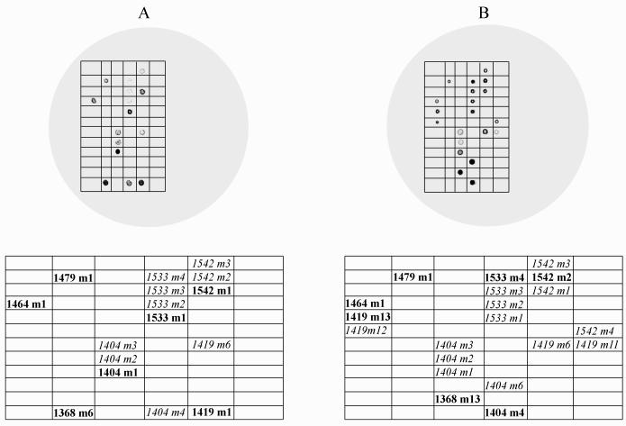
We investigated the application of an oligonucleotide microarray to (i) specifically detect Cryptosporidium spp., (ii) differentiate between closely related C. parvum isolates and Cryptosporidium species, and (iii) differentiate between principle genotypes known to infect humans. A microarray of 68 capture probes targeting seven single-nucleotide polymorphisms (SNPs) within a 190-bp region of the hsp70 gene of Cryptosporidium parvum was constructed. Labeled hsp70 targets were generated by PCR with biotin- or Cy3-labeled primers. Hybridization conditions were optimized for hybridization time, temperature, and salt concentration. Two genotype I C. parvum isolates (TU502 and UG502), two C. parvum genotype II isolates (Iowa and GCH1), and DNAs from 22 non-Cryptosporidium sp. organisms were used to test method specificity. Only DNAs from C. parvum isolates produced labeled amplicons that could be hybridized to and detected on the array. Hybridization patterns between genotypes were visually distinct, but identification of SNPs required statistical analysis of the signal intensity data. The results indicated that correct mismatch discrimination could be achieved for all seven SNPs for the UG502 isolate, five of seven SNPs for the TU502 isolate, and six of seven SNPs for both the Iowa and GCH1 isolates. Even without perfect mismatch discrimination, the microarray method unambiguously distinguished between genotype I and genotype II isolates and demonstrated the potential to differentiate between other isolates and species on a single microarray. This method may provide a powerful new tool for water utilities and public health officials for assessing point and nonpoint source contamination of water supplies.


Figures

FIG. 1.
FIG. 1.
Typical microarray hybridization patterns for C. parvum hsp70 amplicons. (A) Genotype I isolate UG502; (B) genotype II isolate Iowa. The grids below the patterns indicate the hybridized probes and their relative positions on the array. Boldface type indicates the perfectly matched probes for each isolate, and italic type indicates the mismatched probes that also resulted in a positive signal. Images were enhanced for publication by using Adobe Photoshop 6.0, but the underlying data for statistical analysis remained unchanged.
FIG. 2.
FIG. 2.
Detection of SNPs at selected diagnostic positions for genotype I isolates TU502 and UG502. The letters above the probe positions indicate the polymorphic nucleotides for each probe combination. Probes in italics are the perfectly matched probes for genotype I isolates (see Table 1 for the complete probe sequences). Results were based on a minimum of two replicates for each isolate. Error bars represent ±2 standard errors of the mean. All hybridizations were conducted by using the Cy3 labeling strategy and overnight hybridization at 4°C. (A) Positions 1368 and 1371. For clarity, only the probes with the three greatest values are shown. (B) Position 1404. T′ is a single-base-pair mismatch between the genotype II probe, m4, and the probe for C. felis. This polymorphism occurs at position 1398 (numbering based on GenBank accession number AF221535). (C) Position 1533. (D) Position 1542. OD, optical density.
FIG. 3.
FIG. 3.
Detection of SNPs at selected diagnostic positions for genotype II isolates Iowa and GCH1. The letters above the probe positions indicate the polymorphic nucleotides for each probe combination. Probes in italics are the perfectly matched probes for genotype II isolates (see Table 1 for the complete probe sequences). Results were based on a minimum of two replicates for each isolate. Error bars represent ±2 standard errors of the mean. All hybridizations were conducted by using the Cy3 labeling strategy and overnight hybridization at 4°C, except for panel D, where the hybridization temperature was room temperature (ca. 22°C). (A) Positions 1368 and 1371. For clarity, only the probes with the three greatest values are shown. (B) Position 1404. T′ is a single-base-pair mismatch between the perfectly matched probe, m4, and the probe for C. felis. This polymorphism occurs at position 1398 (numbering based on GenBank accession number AF221535). (C) Position 1533. (D) Position 1542. OD, optical density.
FIG. 4.
FIG. 4.
Comparison of hybridization results for genotype I isolates TU502 and UG502 at position 1419. No hybridization signal was detected for 1419 m12 and m13, which are omitted from this graph for clarity. Probe 1419 m1 is the perfectly matched probe for genotype I isolates and the only position of relevance for the genotype I SNP code. Letters above the probes indicate the polymorphic nucleotides at positions 1419 and 1422, respectively. Probes m8 to m13 are outlier probes containing multiple polymorphisms at positions other than and in addition to 1419 and 1422. Results were based on a minimum of two replicates for each isolate. Error bars represent ±2 standard errors of the mean. Hybridizations were conducted by using the Cy3 labeling strategy and overnight hybridization at 4°C. Single-nucleotide mismatch could not be reliably achieved for genotype II isolates when the m13 probe was the perfectly matched probe for these isolates. OD, optical density.

References

    1. Ausubel, F. M., R. Brent, R. E. Kingston, D. D. Moore, J. G. Seidman, J. A. Smith, and K. Struhl (ed.). 1995. Current protocols in molecular biology. John Wiley & Sons, Inc., New York, N.Y.
    1. Call, D. R., D. P. Chandler, and F. Brockman. 2001. Fabrication of DNA microarrays using unmodified oligonucleotide probes. BioTechniques 30:368-379. - PubMed
    1. Clark, D. P. 1999. New insights into human cryptosporidiosis. Clin. Microbiol. Rev. 12:554-563. - PMC - PubMed
    1. Deng, M. Q., and D. O. Cliver. 1998. Differentiation of Cryptosporidium parvum isolates by a simplified randomly amplified polymorphic DNA technique. Appl. Environ. Microbiol. 64:1954-1957. - PMC - PubMed
    1. Drmanac, R., Z. Strezoska, I. Labat, S. Drmanac, and R. Crkvenjakov. 1990. Reliable hybridization of oligonucleotides as short as six nucleotides. DNA Cell Biol. 7:527-534. - PubMed

Publication types

MeSH terms

Substances

LinkOut - more resources