

Inducible gene expression by nonculturable bacteria in milk after pasteurization
- PMID: 11916722
- PMCID: PMC123843
- DOI: 10.1128/AEM.68.4.1988-1993.2002
Inducible gene expression by nonculturable bacteria in milk after pasteurization
Abstract
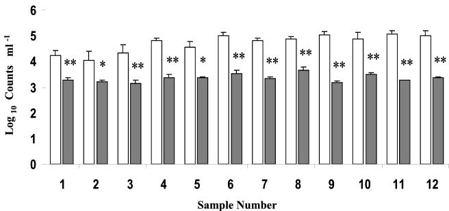
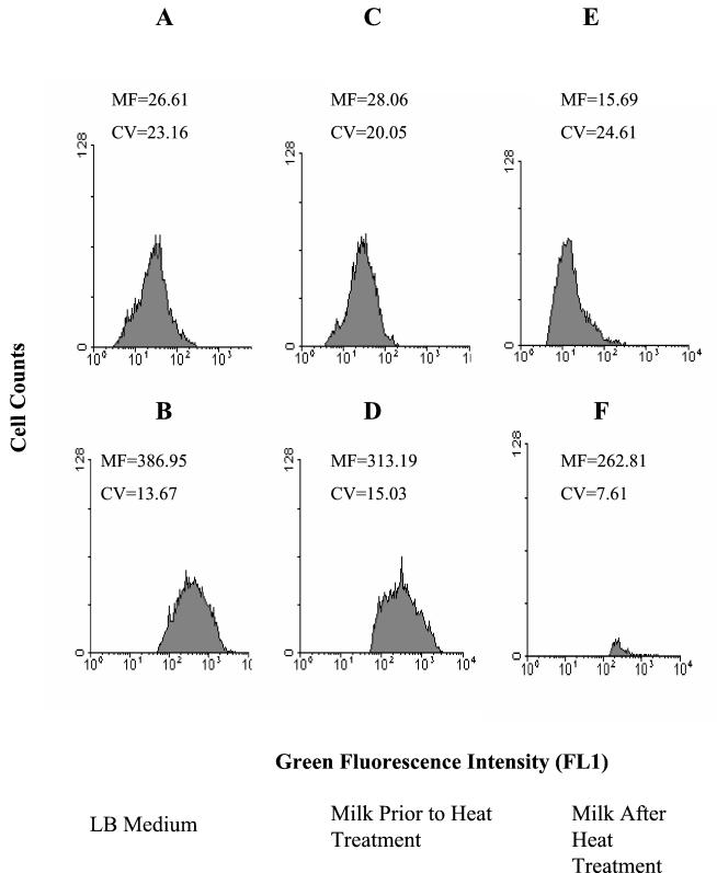
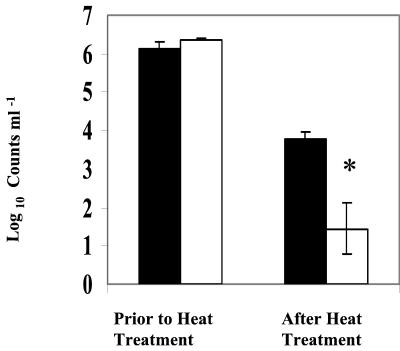
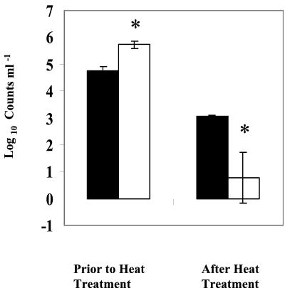
The viability of bacteria in milk after heat treatments was assessed by using three different viability indicators: (i) CFU on plate count agar, (ii) de novo expression of a gfp reporter gene, and (iii) membrane integrity based on propidium iodide exclusion. In commercially available pasteurized milk, direct viable counts, based on dye exclusion, were significantly (P < 0.05) higher than viable cell counts determined from CFU, suggesting that a significant subpopulation of cells in pasteurized milk are viable but nonculturable. Heating milk at 63.5 degrees C for 30 min resulted in a >4-log-unit reduction in the number of CFU of Escherichia coli and Pseudomonas putida that were marked with lac-inducible gfp. However, the reduction in the number of gfp-expressing cells of both organisms under the same conditions was <2.5 log units. These results demonstrate that a substantial portion of cells rendered incapable of forming colonies by heat treatment are metabolically active and are able to transcribe and translate genes de novo.
Figures
References
-
- Auty, M. A. E., G. E. Gardiner, S. J. McBrearty, E. O. O'Sullivan, D. M. Mulvihill, J. K. Collins, G. F. Fitzgerald, C. Stanton, and R. P. Ross. 2001. Direct in situ viability assessment of bacteria in probiotic dairy products using viability staining in conjunction with confocal scanning laser microscopy. Appl. Environ. Microbiol. 67:420-425. - PMC - PubMed
-
- Barer, M. R., and C. R. Harwood. 1999. Bacterial viability and culturability. Adv. Microb. Physiol. 41:93-137. - PubMed
-
- Binderova, E., and D. Rysanek. 1999. Microbial contaminants of milk processed by high-temperature short-time pasteurization. Veterinarni Medicina 44:301-307.
-
- Bloomfield, A. F., G. S. A. B. Stewart, C. E. R. Dodd, I. R. Booth, and E. G. M. Power. 1998. The viable but non-culturable phenomenon explained? Microbiology 14:1-4. - PubMed
-
- Cho, J., and S. Kim. 1999. Viable but non-culturable state of a green fluorescence protein-tagged environmental isolate of Salmonella typhi in groundwater and pond water. FEMS Microbiol. Lett. 170:257-264. - PubMed
MeSH terms
Substances
LinkOut - more resources
Full Text Sources
