. 2002 Apr 1;157(1):103-13.
doi: 10.1083/jcb.200107108. Epub 2002 Mar 26.

The intraflagellar transport protein, IFT88, is essential for vertebrate photoreceptor assembly and maintenance

Affiliations

The intraflagellar transport protein, IFT88, is essential for vertebrate photoreceptor assembly and maintenance

Gregory J Pazour et al. J Cell Biol. .

Abstract

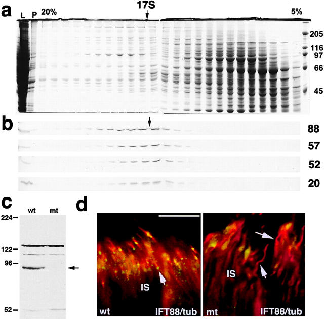
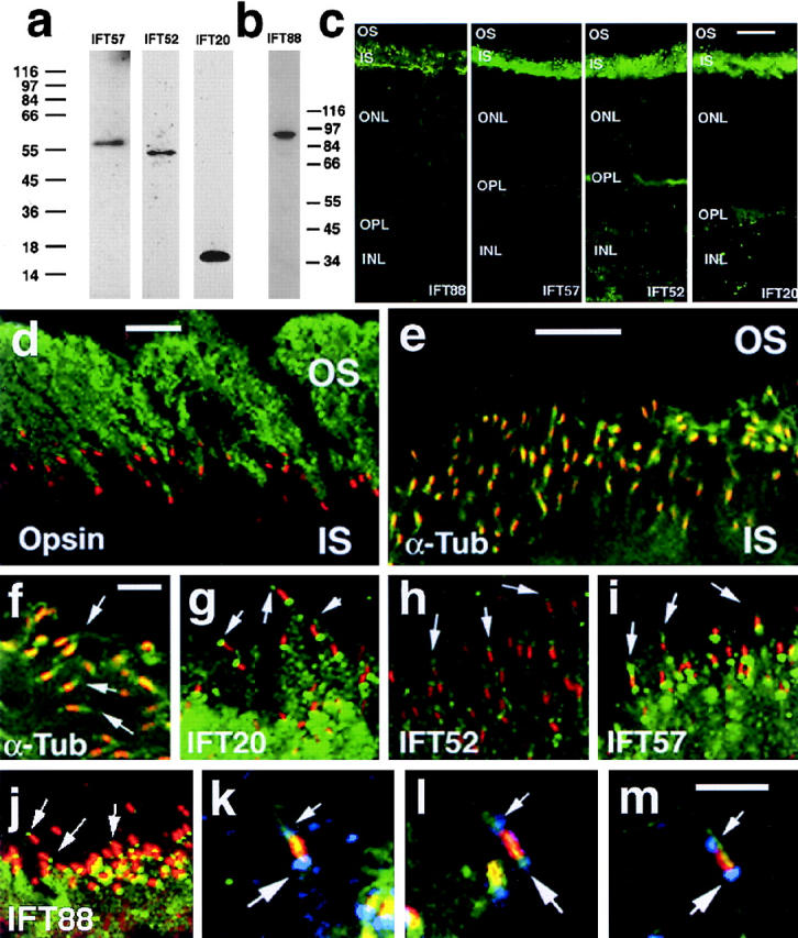
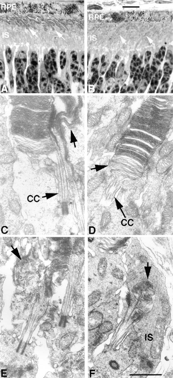
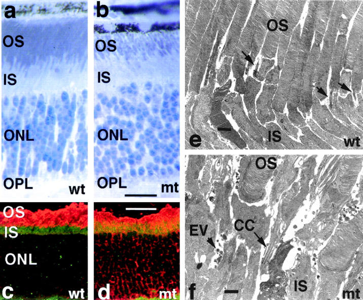
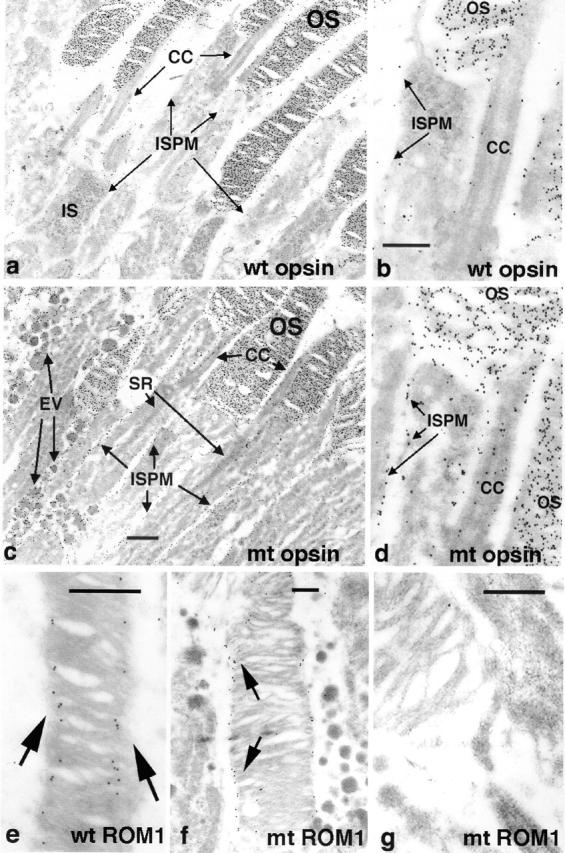
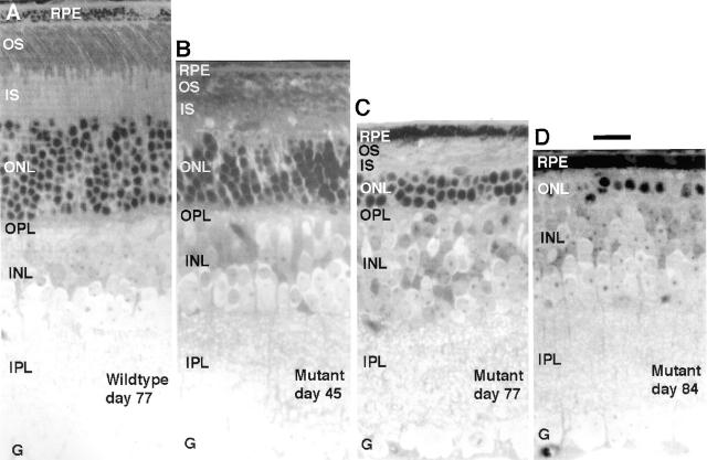
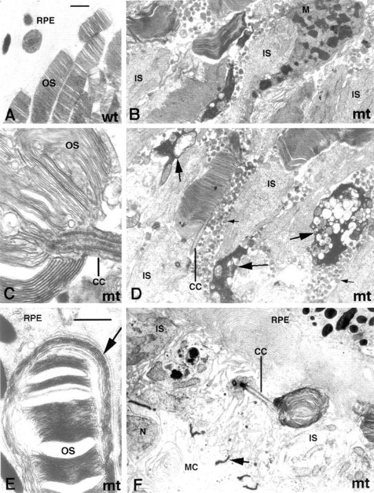
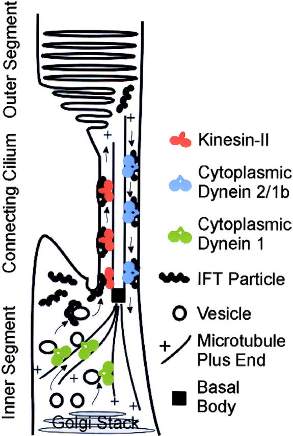
Approximately 10% of the photoreceptor outer segment (OS) is turned over each day, requiring large amounts of lipid and protein to be moved from the inner segment to the OS. Defects in intraphotoreceptor transport can lead to retinal degeneration and blindness. The transport mechanisms are unknown, but because the OS is a modified cilium, intraflagellar transport (IFT) is a candidate mechanism. IFT involves movement of large protein complexes along ciliary microtubules and is required for assembly and maintenance of cilia. We show that IFT particle proteins are localized to photoreceptor connecting cilia. We further find that mice with a mutation in the IFT particle protein gene, Tg737/IFT88, have abnormal OS development and retinal degeneration. Thus, IFT is important for assembly and maintenance of the vertebrate OS.


Figures

Figure 1.
Figure 1.
IFT proteins in mouse testis and retina. (a and b) Sucrose density gradient (5–20%) analysis of a protein extract from mouse testis showing that IFT88, IFT57, IFT52, and IFT20 cosediment at ∼17S. (a) Coomasie blue–stained gels with molecular weight markers in kD indicated on right. (b) Western blots for the four IFT proteins (labeled on the right). Additional abbreviations: L, supernatant protein loaded on gradient; P, pellet from initial protein extraction. (c) Western blot of retinal extracts showing that IFT88 (arrow) is greatly reduced in Tg737−/− (mt) mice as compared with wild type (wt) at p21. (d) Immunofluorescence images showing that IFT88 (green) is found at the ends of the connecting cilia (red, arrows) in wild-type mouse retina but not in Tg737 mutant retina. The connecting cilia were detected by an antibody to acetylated tubulin (red). Bar, 20 μm.
Figure 2.
Figure 2.
IFT particle proteins are found in bovine photoreceptors. (a and b) Western blots demonstrating the presence of IFT particle proteins in a bovine DEPC preparation that is enriched in ciliary axonemes. Antibodies raised against mouse IFT particle proteins recognize single bands of the predicted size (IFT88, 90 kD; IFT57, 57 kD; IFT52, 52 kD; IFT20, 16 kD). Numbers on left (a) and right (b) are molecular weight markers. (c) Panel of confocal immunocytochemistry images of fresh-frozen bovine retina labeled with antibodies to IFT88, IFT57, IFT52, and IFT20 from left to right. IFT particle proteins are localized primarily in the IS. Staining is punctate at the junction of the IS and OS layers where the connecting cilia are located. Staining is also detected in the outer plexiform layer (OPL), where the photoreceptor synaptic terminals are located, and to a lesser extent in the ONL. INL indicates inner nuclear layer. (d–j) Localization of IFT particle proteins to photoreceptor cilia and basal bodies. (d) Bovine OSs are labeled with the B6-30N (green) monoclonal antibody to bovine rhodopsin (Besharse and Wetzel, 1995), and connecting cilia are labeled with the K26 (red) monoclonal antibody. (e and f) Axonemes are labeled with a monoclonal antibody to acetylated α-tubulin (green), and connecting cilia are labeled with K26 (red). Arrows indicate distal ends of cilia. (g–j) Connecting cilia are detected with K26 (red), whereas IFT20 (g), IFT52 (h), IFT57 (i), and IFT88 (j) are detected with affinity-purified rabbit antibodies (green). Arrows indicate distal ends of cilia. (k–m) Triple-labeled images in which labeling with antibodies to IFT57 (blue) is superimposed over labeling of both the connecting cilium (K26, red) and acetylated α-tubulin (green). Small arrows in k–m indicate labeling that is distal to the connecting cilium; the large arrows indicate labeling that is proximal to the connecting cilium. Bars: (c) 20 μm; (d and e) 10 μm; (f) 5 μm; (m) 2 μm; (g–j) refer to e; (k and l) refer to m.
Figure 3.
Figure 3.
Rod cell OS is abnormal in Tg737 mutant mice at p10. (A) Typical wild-type retina showing the extent of OS development (arrows) at p10. (B) Tg737 mutant retina at p10 showing smaller and less dense OSs (arrows) than seen in wild type. RPE indicates retinal pigment epithelium. (C and D) Examples of OSs in wild-type animals at p10. In C, the arrow indicates an abnormal OS adjacent to a normal one. Such atypical OSs are only occasionally observed in wild-type mice. In D, the arrow indicates discs in the process of being formed adjacent to the connecting cilium (CC). (E and F) Typical examples of aberrant OSs in Tg737 mutant mice at p10. Disrupted discs (E, arrow) and OSs extending into their own ISs (F, arrow) are widespread in Tg737 mutant retinas at p10. Bars: (A and B) 10 μm; (C–F) 1 μm.
Figure 4.
Figure 4.
Rod cell OSs are abnormal and opsin is mislocalized in Tg737 mutant mice at p21. (a and b) Toluidine-blue–stained sections illustrating that mutant OSs (b) are less organized and less densely packed than those in similar sections of wild type (a). (c and d) Immunocytochemistry of rod opsin (red) and IFT88 (green) in frozen sections of wild-type (c) and Tg737 mutant (d) retina at p21. Both wild-type and mutant animals show strong opsin staining of the rod OS, but opsin is also detected in IS and around nuclei (ONL) in the mutant. IFT88 is present in the IS (c) but diminished in the mutant (d). (e and f) Electron micrographs of the junction between the IS and OS layers illustrating that mutant (f) photoreceptors are less uniform in structure and organization than wild-type photoreceptors (e) and accumulate extracellular vesicles (EV). Other abbreviations are the same as in figures 2 and 3. Bars: (a and b) 20 μm; (c and d) 40 μm; (e and f) 1 μm.
Figure 7.
Figure 7.
EM level immunocytochemical localization of rod opsin and ROM1 in OSs of Tg737 mutant mice. (a and b) Distribution of opsin in OSs of wild-type retinas at p21. Immunoreactivity is abundant in OSs, but very low levels are detected in the IS plasma membrane (ISPM) and connecting cilium (CC). (c and d) Distribution of opsin in OSs of Tg737 mutant retinas at p21. Opsin exhibits an abundance in mutant OSs similar to that in wild type, but is found at much higher levels compared with wild type in the IS plasma membrane and connecting cilium. In addition, opsin is present in the extracellular vesicles (EV) that are not found in wild-type animals. (e–g) Distribution of ROM1 in wild-type (e) and Tg737 (f and g) photoreceptors. ROM1 (arrows) is localized at disc edges and at incisures in wild-type animals. A similar pattern is detected in those Tg737 mutant rods that exhibit organized discs (f), but ROM1 appears to be much less abundant in OS with disorganized discs (g). Bars: (b and c) 1 μm; (e, f, and g) 0.5 μm; (a) refer to c; (d) refer to b.
Figure 5.
Figure 5.
Light microscopic images demonstrating progressive loss of photoreceptors in Tg737 mutant mice. (A) Full thickness section of typical wild-type retina at p77. (B–D) Full thickness views of retinas from Tg737 mutant mice at p45, p77, and p84, showing loss of nuclei from the ONL and progressive thinning of the IS and OS layers. All images are from 1-μm-thick plastic sections stained with toluidine blue. Other abbreviations: G, ganglion cell layer; INL, inner nuclear layer; IPL, inner plexiform layer; OPL, outer plexiform layer. Bar, 20 μm.
Figure 6.
Figure 6.
EM images showing aberrant photoreceptor organization in Tg737 homozygous mutants at p45 and p77. (A) An image of typical well-organized OSs from wild-type animals. (B–D) Tg737 mutant mice at p45. Abundant extracellular vesicles (D, small arrows) with an amorphous interior accumulate between living photoreceptors; this is never seen in wild-type animals. Condensed, dying photoreceptors are frequently seen throughout the photoreceptor layer (D, large arrows), and macrophages (B, M) are sometimes seen among photoreceptors. (E and F) Late stage of photoreceptor loss at p77. In E, a single aberrant OS adjacent to the pigment epithelium (RPE) is seen with relatively normal discs surrounded by exceptionally large disc membranes. In F, a single underdeveloped OS with whorls of disc membranes and its connecting cilium (CC) are seen adjacent to the RPE. Note that Mueller cell bodies (MC) and the junctional complexes between them (F, arrow), which normally are separated from the RPE by the full thickness of the photoreceptor IS and OS layers, are seen in the same field of view as the RPE in F. Bars: (A and E) 1 μm; (B, D, and F) refer A; (C) refer to E.
Figure 8.
Figure 8.
Model of photoreceptor cell IFT. Cytoplasmic dynein 1 transports vesicles from the Golgi stack to the base of the connecting cilium. IFT particles associate with the vesicles and the vesicles fuse with the ciliary or plasma membrane at the base of the connecting cilium. The IFT particles with attached cargo are then transported through the connecting cilium by kinesin-II. At the distal end of the connecting cilium, the IFT particles dissociate from their cargo, the membrane is organized into disks, and the IFT particles are picked up by cytoplasmic dynein 1b/2 to be returned to the cell body.

References

    1. Anderson, R.E., M.B. Maude, P.A. Kelleher, T.M. Maida, and S.F. Basinger. 1980. Metabolism of phosphatidylcholine in the frog retina. Biochim. Biophys. Acta. 620:212–226. - PubMed
    1. Beech, P.L., K. Pagh-Roehl, Y. Noda, N. Hirokawa, B. Burnside, and J.L. Rosenbaum. 1996. Localization of kinesin superfamily proteins to the connecting cilium of fish photoreceptors. J. Cell Sci. 109:889–897. - PubMed
    1. Besharse, J.C., and C.J. Horst. 1990. The photoreceptor connecting cilium. A model for the transition zone. Ciliary and Flagellar Membranes. R.A. Bloodgood, editor. Plenum Publishing Corp, New York. 389–417.
    1. Besharse, J.C., and M.G. Wetzel. 1995. Immunocytochemical localization of opsin in rod photoreceptors during periods of rapid disc assembly. J. Neurocytol. 24:371–388. - PubMed
    1. Blanks, J.C., and C. Spee. 1992. Retinal degeneration in the pcd/pcd mutant mouse: accumulation of spherules in the interphotoreceptor space. Exp. Eye Res. 54:637–644. - PubMed

Publication types

MeSH terms