. 2002 Apr 1;157(1):91-101.
doi: 10.1083/jcb.200112080. Epub 2002 Mar 26.

Mammalian class E vps proteins recognize ubiquitin and act in the removal of endosomal protein-ubiquitin conjugates

Affiliations

Mammalian class E vps proteins recognize ubiquitin and act in the removal of endosomal protein-ubiquitin conjugates

Naomi Bishop et al. J Cell Biol. .

Abstract

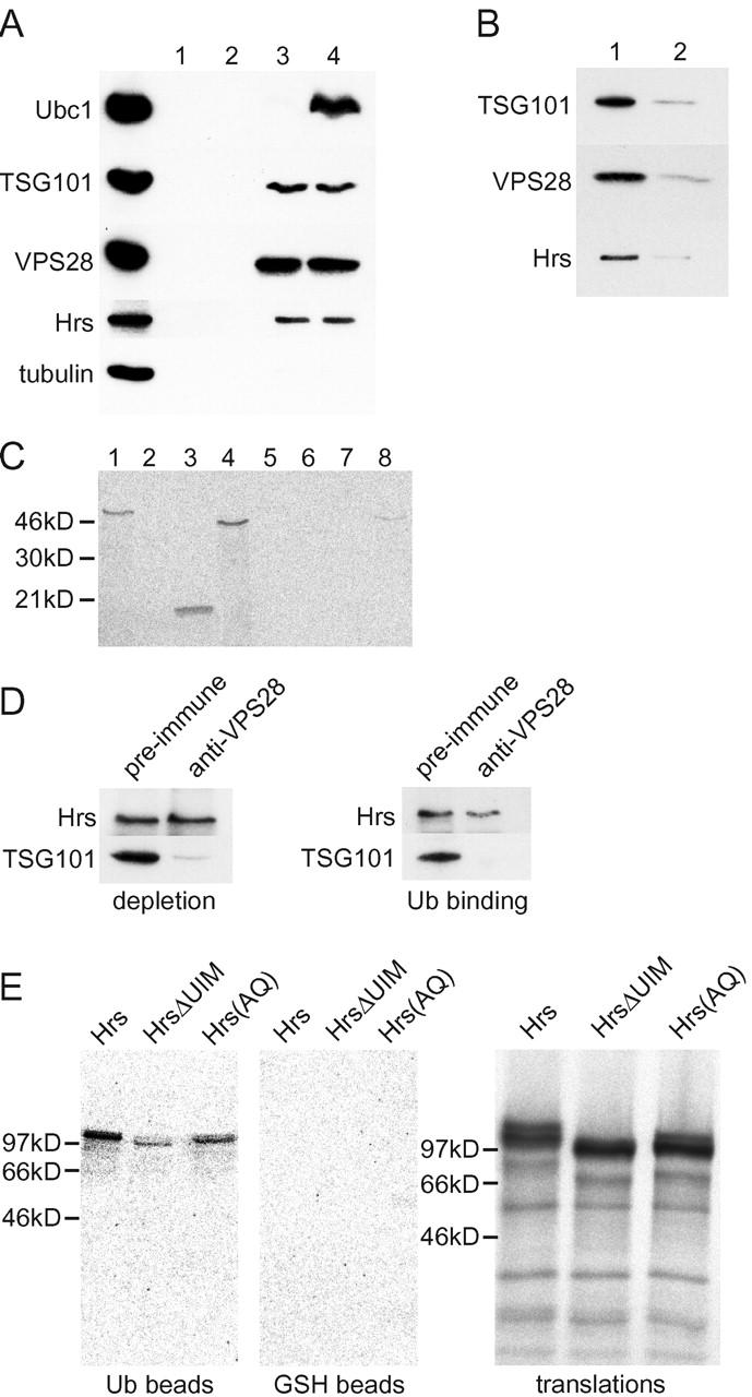
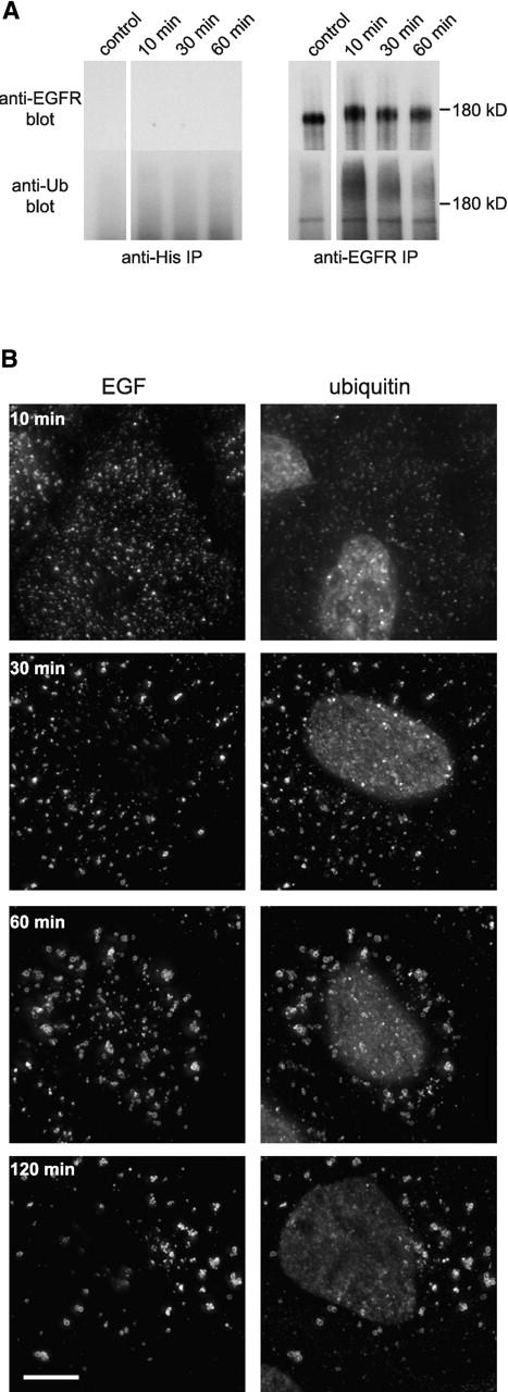
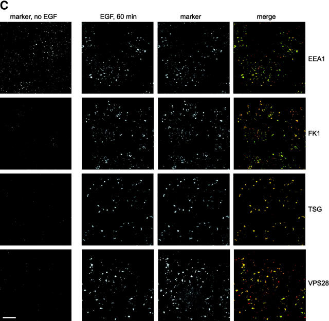
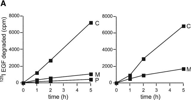
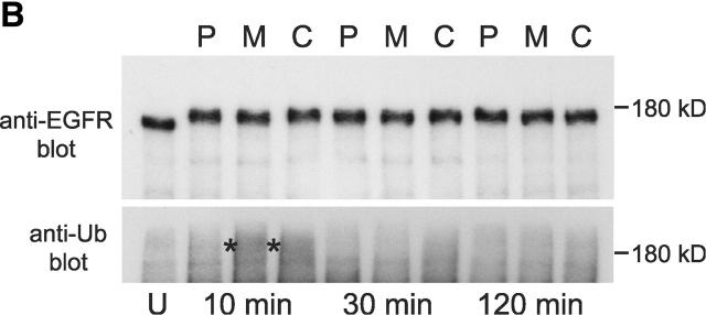
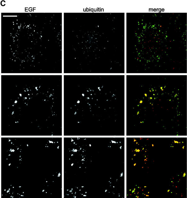
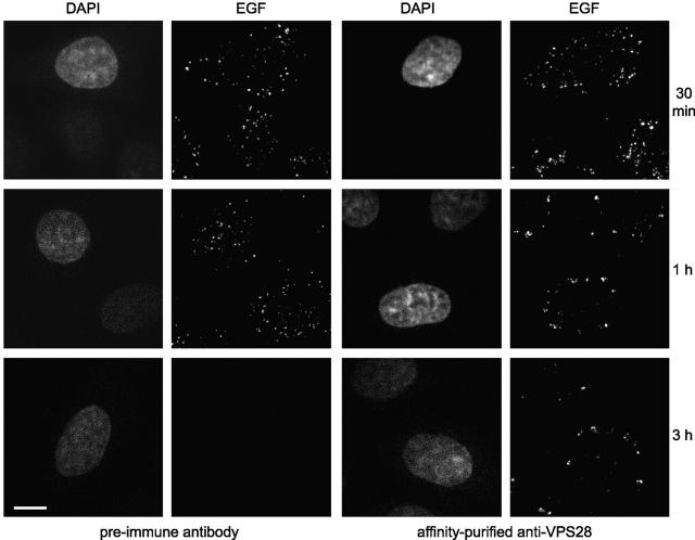
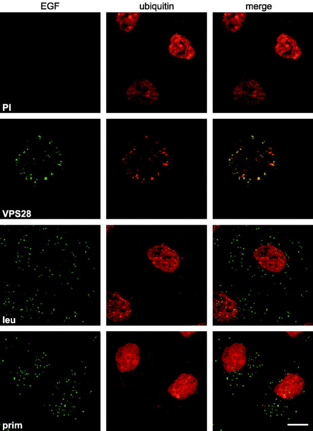
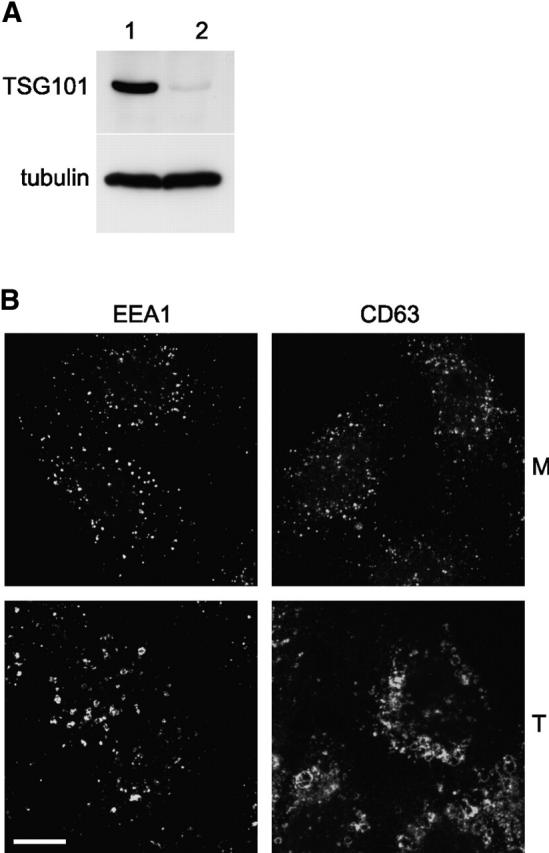
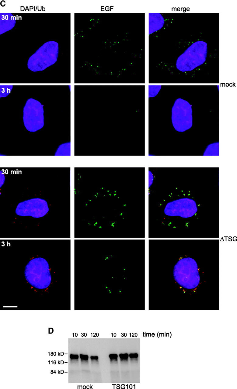
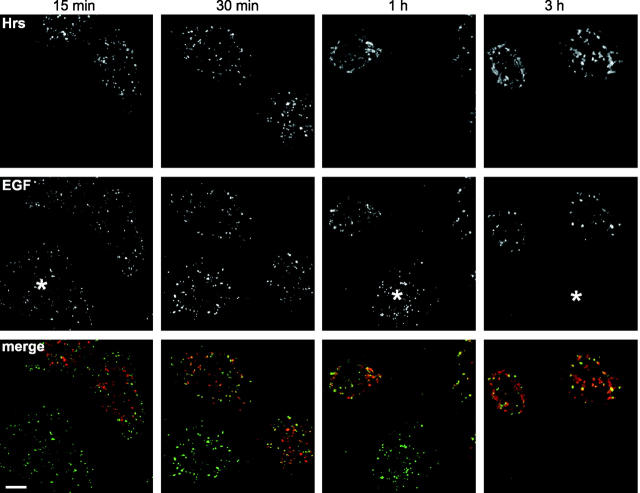
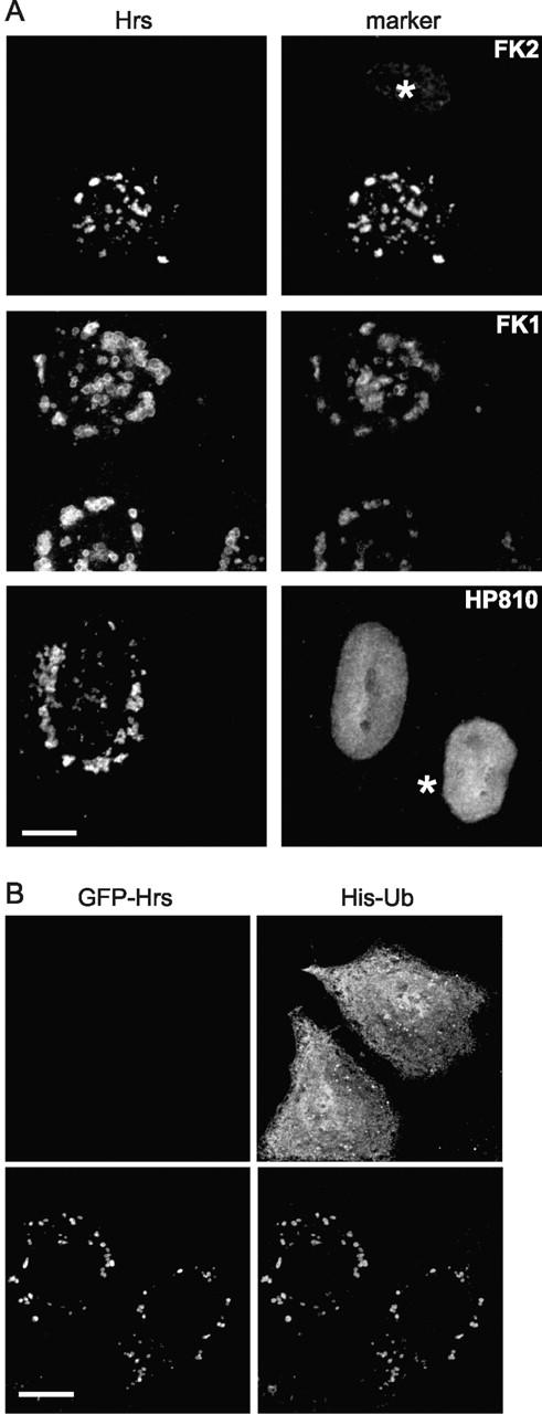
There is increasing evidence that ubiquitination of receptors provides an important endosomal sorting signal. Here we report that mammalian class E vacuolar protein-sorting (vps) proteins recognize ubiquitin. Both tumor susceptibility gene 101 (TSG101)/human VPS (hVPS)28 and hepatocyte growth factor receptor substrate (Hrs) cytosolic complexes bind ubiquitin-agarose. TSG101 and hVPS28 are localized to endosomes that contain internalized EGF receptor and label strongly for ubiquitinated proteins. Microinjection of anti-hVPS28 specifically retards EGF degradation and leads to endosomal accumulation of ubiquitin-protein conjugates. Likewise, depletion of TSG101 impairs EGF trafficking and causes dramatic relocalization of ubiquitin to endocytic compartments. Similar defects are found in cells overexpressing Hrs, further emphasizing the links between class E protein function, receptor trafficking, and endosomal ubiquitination.


Figures

Figure 1.
Figure 1.
Mammalian class E vps proteins bind ubiquitin. (A) ATP-depleted HeLa cytosol was incubated with GSH- (lanes 1 and 2) or ubiquitin-agarose (lanes 3 and 4) with (lanes 1 and 3) or without (lanes 2 and 4) 1 mM Mg-ATP. Bound fractions were analyzed by Western blotting. 25% of the input was also blotted (T). (B) Cytosol was incubated with ubiquitin-agarose after preincubating for 15 min without (lane 1) or with (lane 2) 1 mg soluble ubiquitin. Bound fractions were analyzed as above. (C) Equal counts of in vitro–translated TSG101 (lanes 1 and 5), TSG101ΔUbc (lanes 2 and 6), the Ubc domain alone (lanes 3 and 7), or TSG101ΔHx (lanes 4 and 8) were incubated with ubiquitin- (lanes 1–4) or GSH-agarose (lanes 5–8). Bound fractions were analyzed by PAGE and phosphorimaging. (D) HeLa cytosol was immunodepleted using either preimmune or anti-hVPS28 IgG. The resulting cytosols were Western blotted for Hrs and TSG101 (left). Parallel samples were incubated with ubiquitin-agarose, and bound material was Western blotted (right). (E) Equal counts of in vitro–translated Hrs, HrsΔUIM, and Hrs(AQ) were incubated with GSH- or ubiquitin-agarose (left). Translation products, run on a separate gel, are also shown. Samples were analyzed as above.
Figure 2.
Figure 2.
TSG101 and hVPS28 are localized to ubiquitin-rich EGF-positive endosomes. (A) A431 cells were starved overnight in serum-free medium and either left untreated (no EGF) or incubated with 0.4 μg/ml EGF at 4°C followed by the indicated time at 37°C. Cell lysates were immunoprecipitated with control (left) or anti-EGFR antibody (right). Bound fractions were Western blotted with anti-EGFR (top) or antiubiquitin (FK2; bottom). The band detected in the FK2 blot running just below the EGFR is an artifact, since it is found in EGFR immunoprecipitates from cells lacking EGFR (unpublished data). (B) Oregon green EGF was bound to A431 cells and internalized as indicated. Cells were chilled, incubated with saponin before fixation, and then analyzed for EGF content (left) and stained with FK2 antibody (right). (C) A431 cells were either left untreated (left) or had Oregon green EGF internalized for 60 min (right). Cells were chilled and incubated with saponin before fixation and then analyzed for EGF (green) or stained (red) for the following markers: EEA1, polyubiquitin–protein conjugates (FK1), TSG101, or hVPS28.
Figure 2.
Figure 2.
TSG101 and hVPS28 are localized to ubiquitin-rich EGF-positive endosomes. (A) A431 cells were starved overnight in serum-free medium and either left untreated (no EGF) or incubated with 0.4 μg/ml EGF at 4°C followed by the indicated time at 37°C. Cell lysates were immunoprecipitated with control (left) or anti-EGFR antibody (right). Bound fractions were Western blotted with anti-EGFR (top) or antiubiquitin (FK2; bottom). The band detected in the FK2 blot running just below the EGFR is an artifact, since it is found in EGFR immunoprecipitates from cells lacking EGFR (unpublished data). (B) Oregon green EGF was bound to A431 cells and internalized as indicated. Cells were chilled, incubated with saponin before fixation, and then analyzed for EGF content (left) and stained with FK2 antibody (right). (C) A431 cells were either left untreated (left) or had Oregon green EGF internalized for 60 min (right). Cells were chilled and incubated with saponin before fixation and then analyzed for EGF (green) or stained (red) for the following markers: EEA1, polyubiquitin–protein conjugates (FK1), TSG101, or hVPS28.
Figure 3.
Figure 3.
Inhibition of proteasome function inhibits EGF degradation and prevents deubiquitination of endosomal proteins. (A, left) 125I EGF was bound and internalized into A431 cells in the absence (control; C) or presence (M) of 5 μM MG132 or with MG132 in cells which had also been preincubated for 3 h with 5 μM MG132 (P). After the indicated times of internalization, medium was removed and assayed for degraded 125I EGF. (A, right) 125I EGF was bound and internalized into untreated A431 cells (C) or into cells to which MG132 was added 30 min after internalization had begun (M). Samples were assayed for EGF degradation as above. All values are means of triplicate determinations ± SEM. (B) A431 Cells were incubated with 0.4 μg/ml EGF at 4°C followed by 10, 30, or 120 min at 37°C as indicated. Cells were incubated with 5 μM MG132 for 3 h before and during internalization (P), incubated with 5 μM MG132 during internalization only (M), or otherwise untreated (C). Cell lysates were immunoprecipitated with anti-EGFR antibody, and bound fractions were Western blotted with anti-EGFR (top) or antiubiquitin (FK2; bottom). The samples containing peak FK2 labeling are indicated by asterisks. An immunoprecipitate from untreated cells is also shown (U). (C) Oregon green EGF was bound to A431 cells and internalized. Cells were chilled, incubated with saponin before fixation, and then analyzed for EGF content (green) and stained with FK2 antibody (red). (Top) Cells preincubated for 3 h with MG132 and then EGF internalized for 1 h with MG132. (Middle) EGF internalized for 1 h with MG132. (Bottom) EGF internalized for 30 min followed by a further 4.5 h with MG132.
Figure 3.
Figure 3.
Inhibition of proteasome function inhibits EGF degradation and prevents deubiquitination of endosomal proteins. (A, left) 125I EGF was bound and internalized into A431 cells in the absence (control; C) or presence (M) of 5 μM MG132 or with MG132 in cells which had also been preincubated for 3 h with 5 μM MG132 (P). After the indicated times of internalization, medium was removed and assayed for degraded 125I EGF. (A, right) 125I EGF was bound and internalized into untreated A431 cells (C) or into cells to which MG132 was added 30 min after internalization had begun (M). Samples were assayed for EGF degradation as above. All values are means of triplicate determinations ± SEM. (B) A431 Cells were incubated with 0.4 μg/ml EGF at 4°C followed by 10, 30, or 120 min at 37°C as indicated. Cells were incubated with 5 μM MG132 for 3 h before and during internalization (P), incubated with 5 μM MG132 during internalization only (M), or otherwise untreated (C). Cell lysates were immunoprecipitated with anti-EGFR antibody, and bound fractions were Western blotted with anti-EGFR (top) or antiubiquitin (FK2; bottom). The samples containing peak FK2 labeling are indicated by asterisks. An immunoprecipitate from untreated cells is also shown (U). (C) Oregon green EGF was bound to A431 cells and internalized. Cells were chilled, incubated with saponin before fixation, and then analyzed for EGF content (green) and stained with FK2 antibody (red). (Top) Cells preincubated for 3 h with MG132 and then EGF internalized for 1 h with MG132. (Middle) EGF internalized for 1 h with MG132. (Bottom) EGF internalized for 30 min followed by a further 4.5 h with MG132.
Figure 4.
Figure 4.
Anti-hVPS28 inhibits EGF degradation. HeLa cells were microinjected with preimmune IgG (left) or anti-hVPS28 (right). Texas red EGF was internalized as indicated, and cells were examined for EGF. Injected cells were detected by DAPI staining. Confocal settings were identical for all samples.
Figure 5.
Figure 5.
Anti-hVPS28 causes ubiquitinated proteins to accumulate on endosomes. HeLa cells were microinjected with preimmune IgG (PI) or anti-hVPS28 antibody (VPS28). Oregon green EGF was internalized for 3 h. Alternatively, Oregon green EGF was bound and internalized into cells treated with either 100 μM leupeptin (leu) or 0.3 mM primaquine (prim). Cells were permeabilized with saponin, fixed, visualized for EGF (EGF), and stained with FK2 monoclonal antibody. Microinjected cells were identified by DAPI staining (unpublished data). Confocal settings were identical for all EGF labeling but were altered for ubiquitin staining to prevent saturation.
Figure 6.
Figure 6.
Depletion of TSG101 affects EGF trafficking and endosomal ubiquitination. (A) HeLa cells were mock transfected (lane 1) or transfected with siRNA against Tsg101 (lane 2). Cell extracts were Western blotted for TSG101 (top) or tubulin as a control (bottom). (B) HeLa cells were mock transfected (M; top) or transfected with siRNA against Tsg101 (T; bottom) and stained for EEA1 or CD63 as indicated. (C). Oregon green EGF was internalized for the indicated times into mock-transfected HeLa cells (top) or cells transfected with siRNA against Tsg101 (bottom). Cells were analyzed for EGF (green) and stained with DAPI (blue) and with FK2 monoclonal antibody followed by Texas red 2° antibody (red). (D). EGF was bound and internalized into mock-transfected HeLa cells (left) or cells transfected with siRNA against Tsg101 (right). Cell lysates were made after the indicated times of EGF internalization and Western blotted for EGFR.
Figure 6.
Figure 6.
Depletion of TSG101 affects EGF trafficking and endosomal ubiquitination. (A) HeLa cells were mock transfected (lane 1) or transfected with siRNA against Tsg101 (lane 2). Cell extracts were Western blotted for TSG101 (top) or tubulin as a control (bottom). (B) HeLa cells were mock transfected (M; top) or transfected with siRNA against Tsg101 (T; bottom) and stained for EEA1 or CD63 as indicated. (C). Oregon green EGF was internalized for the indicated times into mock-transfected HeLa cells (top) or cells transfected with siRNA against Tsg101 (bottom). Cells were analyzed for EGF (green) and stained with DAPI (blue) and with FK2 monoclonal antibody followed by Texas red 2° antibody (red). (D). EGF was bound and internalized into mock-transfected HeLa cells (left) or cells transfected with siRNA against Tsg101 (right). Cell lysates were made after the indicated times of EGF internalization and Western blotted for EGFR.
Figure 7.
Figure 7.
Hrs expression inhibits EGF degradation. HeLa cells were transfected with Hrs. Oregon green EGF was bound and internalized for the indicated times. Cells were visualized for EGF (green) and stained with anti-Hrs IgG (red). Untransfected cells are indicated by an asterisk.
Figure 8.
Figure 8.
Hrs expression causes relocalization of ubiquitinated proteins. (A) HeLa cells were transfected with untagged Hrs, permeabilized in saponin, fixed, and stained for Hrs (left) and with FK2 or FK1 antiubiquitin–protein conjugates, or with HP810 antiproteasome as indicated (right). Untransfected cells are denoted by an asterisk. (B) HeLa cells were transfected with His6-tagged ubiquitin alone (top) or with His6-tagged ubiquitin and EGFP-Hrs (bottom). Cells were fixed directly in methanol without prior permeabilization and visualized for Hrs (left) and stained using anti-His antibody (right).

References

    1. Amerik, A.Y., J. Nowak, S. Swaminathan, and M. Hochstrasser. 2000. The Doa4 deubiquitinating enzyme is functionally linked to the vacuolar protein-sorting and endocytic pathways. Mol. Biol. Cell. 11:3365–3380. - PMC - PubMed
    1. Babst, M., B. Wendland, E.J. Estapa, and S.D. Emr. 1998. The Vps4p AAA ATPase regulates membrane association of a Vps protein complex required for normal endosome function. EMBO J. 17:2982–2993. - PMC - PubMed
    1. Babst, M., G. Odorizzi, E.J. Estepa, and S.D. Emr. 2000. Mammalian tumor susceptibility gene 101 (TSG101) and the yeast homologue, Vps23p, both function in late endosomal trafficking. Traffic. 1:248–258. - PubMed
    1. Bishop, N., and P.G. Woodman. 2000. ATPase-defective mammalian VPS4 localizes to aberrant endosomes and impairs cholesterol trafficking. Mol. Biol. Cell. 11:227–239. - PMC - PubMed
    1. Bishop, N., and P. Woodman. 2001. Tsg101/mammalian vps23 and mammalian vps28 interact directly and are recruited to vps4-induced endosomes. J. Biol. Chem. 276:11735–11742. - PubMed

Publication types

MeSH terms