. 2002 Apr 1;157(1):79-89.
doi: 10.1083/jcb.200112098. Epub 2002 Mar 26.

A cycle of Vam7p release from and PtdIns 3-P-dependent rebinding to the yeast vacuole is required for homotypic vacuole fusion

Affiliations

A cycle of Vam7p release from and PtdIns 3-P-dependent rebinding to the yeast vacuole is required for homotypic vacuole fusion

Christine Boeddinghaus et al. J Cell Biol. .

Abstract

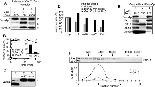
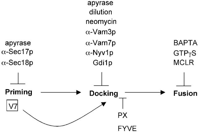
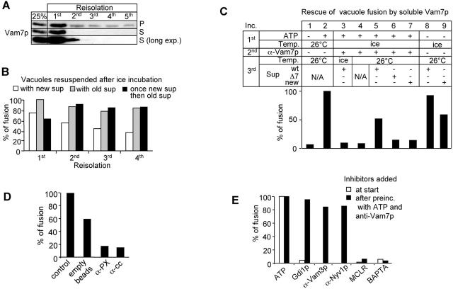
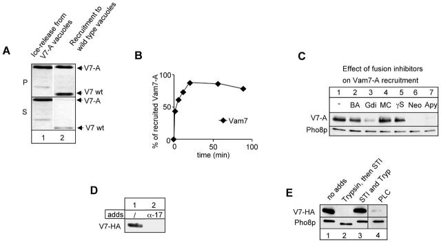
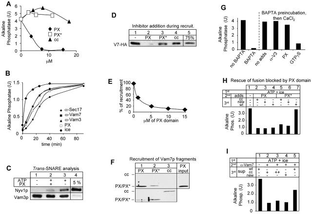
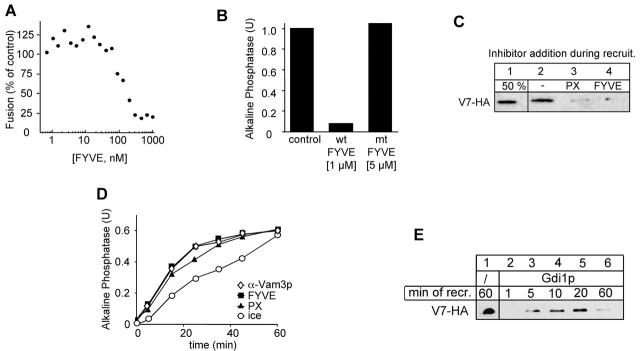
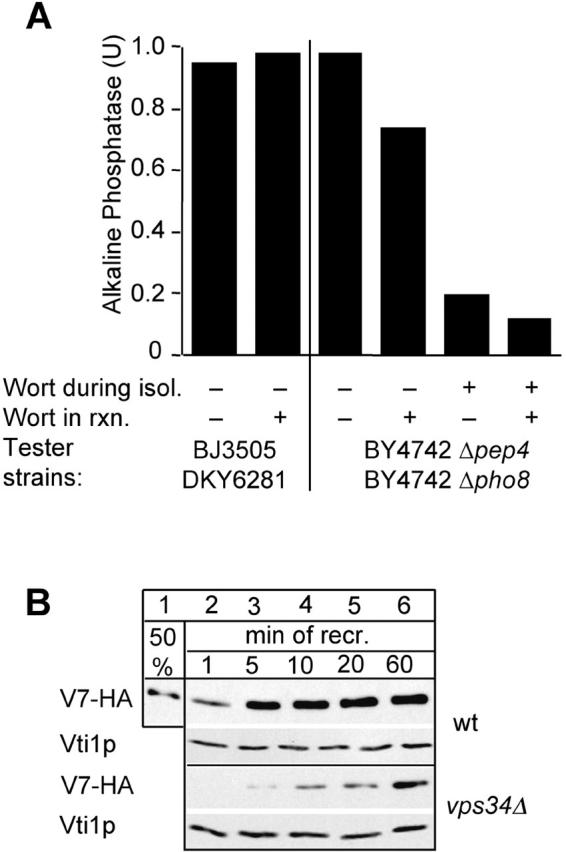
Vacuole fusion requires a coordinated cascade of priming, docking, and fusion. SNARE proteins have been implicated in the fusion itself, although their precise role in the cascade remains unclear. We now report that the vacuolar SNAP-23 homologue Vam7p is a mobile element of the SNARE complex, which moves from an initial association with the cis-SNARE complex via a soluble intermediate to the docking site. Soluble Vam7p is specifically recruited to vacuoles and can rescue a fusion reaction poisoned with antibodies to Vam7p. Both the recombinant Vam7p PX domain and a FYVE domain construct of human Hrs block the recruitment of Vam7p and vacuole fusion, demonstrating that phosphatidylinositol 3-phosphate is a primary receptor of Vam7p on vacuoles. We propose that the Vam7p cycle is linked to the availability of a lipid domain on yeast vacuoles, which is essential for coordinating the fusion reaction prior to and beyond docking.


Figures

Figure 1.
Figure 1.
Priming-dependent release of Vam7p on ice. (A) Vam7p is released from the vacuoles in an ATP-dependent reaction. BJ3505 vacuoles (12 μg) were incubated in a 60-μl reaction for 60 min on ice or at 26°C in reaction buffer containing His6-Sec18p, CoA (10 μM), and ATP as indicated. Then vacuoles were separated from the supernatant (5 min, 20,000 g, 4°C), and the reaction supernatant was precipitated by addition of TCA (13% [vol/vol]) (Ungermann et al., 1998a). Vacuole pellets and precipitated proteins from the supernatant were solubilized in sample buffer and analyzed by SDS-PAGE and immunoblotting. Immunoblots were decorated with indicated antibodies. (B) Release of Vam7p and Sec17p on ice is immediate. BJ3505 vacuoles (12 μg) were incubated on ice, separated into vacuole and supernatant fraction, and processed as in A. Immunoblots of the supernatant fractions were quantified and plotted versus time. The inset shows a quantification of Vam7p in the reaction supernatant after a 60-min incubation on ice or at 26°C. (C) Priming inhibitors block Vam7p release. BJ3505 VAM7-HA vacuoles (12 μg) were incubated in the presence of ATP and IgGs to Sec17p and Sec18p where indicated. Pellet and supernatant fractions were analyzed with HA antibodies to detect Vam7p. HA-tagged Vam7p was used to avoid interference with the added IgGs during immunoblot decoration. (D) Incubation with ATP on ice allows bypass of priming. BJ3505 and DKY6281 vacuoles (6 μg) were incubated in the presence of ATP at the indicated temperature. Inhibitors (α-Sec17p, α-Sec18p [200 μg/ml], α-Vam3p [50 μg/ml], and Gdi1p [300 μg/ml]) were added to the reactions either right away (white bars), after a 30-min ice incubation (gray bars), or after a 30-min incubation at 26°C. Each reaction was then incubated for an additional 60 min at 26°C and then assayed for alkaline phosphatase activity. (E) Vam7p is released from the cis-SNARE complex. BJ3505 vacuoles (36 μg) were incubated in a 180-μl reaction on ice for 10 min. Vacuoles were reisolated (5 min, 20,000 g, 4°C) and solubilized in lysis buffer (150 mM KCl, 0.5% Triton X-100, 20 mM Hepes/KOH, pH 7.4, 1xPIC, and 1 mM PMSF) for 10 min at 4°C and centrifuged (20,000 g, 10 min, 4°C). The soluble detergent extract was added to protein A–Sepharose–coupled antibodies to Vam3p (Ungermann et al., 1998a) and incubated for 2 h at 4°C on a nutator. Reisolated beads were washed once with lysis buffer and once with lysis buffer containing 500 mM KCl. Bound proteins were eluted from the beads with 1 ml of 0.1 M glycine, pH 2.6. The eluate was TCA precipitated and analyzed by SDS-PAGE, Western transfer and immunoblotting with antibodies against the indicated proteins. (F) Released Vam7p is a monomer. BJ 3505 vacuoles (60 μg) were incubated in the presence of ATP on ice or at 26°C as described in the legend to Fig. 1 A. The supernatant was loaded onto a continuous glycerol gradient (10–30% in PS buffer supplemented with 150 mM KCl). The samples were centrifuged at 40,000 rpm in a SW40 rotor for 18 h at 4°C. Fifteen 500-μl fractions were collected, TCA precipitated, and analyzed by SDS-PAGE and immunoblot as before.
Figure 7.
Figure 7.
Schematic overview of Vam7p rebinding to vacuoles. The three basic stages of the vacuole fusion reaction and the respective inhibitors are shown. V7, Vam7p; PX, PX domain of Vam7p; FYVE, GST2xFYVE of Hrs.
Figure 2.
Figure 2.
Released Vam7p is functional. (A) Complete Vam7p release during consecutive ice incubations. BJ3505 vacuoles (12 μg) were incubated in the presence of ATP on ice. After 30 min, the reaction was centrifuged (5 min, 20,000 g, 4°C), supernatant was removed, and a fresh reaction mix was added to the vacuoles. This procedure was repeated four times. The supernatant and the pellet were analyzed by immunoblotting with antibodies to Vam7p. Loss of Vam7p in the supernatant in the second reaction is due to the instability of released Vam7p (see text). (B) Removal of the supernatant coincides with loss of fusion activity. A fusion reaction containing the two tester strains was incubated as in A. After each incubation, vacuoles were reisolated (5 min, 20,000 g, 4°C) and resuspended either in new reaction mix containing Sec18p and CoA (white bars), the same supernatant (gray bars) to monitor loss of fusion due to centrifugation, or in the Vam7-containing supernatant removed in the first incubation (black bars). All samples were then incubated for 60 min at 26°C before assaying for fusion activity. (C) Fusion of vacuoles poisoned with anti-Vam7p. Fusion reactions containing both tester strains were incubated in a first incubation on ice or at 26°C for 30 min. Vacuoles were then reisolated (20,000 g, 5 min, 4°C), incubated for 5 min with or without anti-Vam7p on ice, and reisolated. Supernatant of a parallel reaction (30 min on ice in the presence of ATP) containing wild-type vacuoles or vam7Δ vacuoles was added to the pretreated vacuoles, incubated for 60 min at 26°C, and assayed for alkaline phosphatase activity. (D) Depletion of Vam7p from the supernatant blocks fusion. Priming on ice was done two times as described in A. The supernatant containing released Vam7p was incubated for 30 min at 4°C with protein A–Sepharose containing nonimmune IgG, anti-Vam7 (1–122) (α-PX), or anti-Vam7 (165–316) (α-cc). Then, the cleared supernatant was added to primed vacuoles as in A. (E) Soluble Vam7p can rescue fusion at a stage that is insensitive to docking inhibitors. Inhibitors were added to a fusion reaction either from the beginning (white bars) or after pretreatment with anti-Vam7p (black bars) as described in C. Fusion reactions were then incubated for 60 min at 26°C, and fusion activity was determined.
Figure 3.
Figure 3.
Recruitment of soluble Vam7p to vacuoles. (A) Wild-type vacuoles (BJ3505) and Vam7-A vacuoles were incubated in the presence of ATP for 30 min on ice. Vacuoles were reisolated (20,000 g, 5 min, 4°C), and wild-type vacuoles were resuspended in supernatant removed from Vam7-A vacuoles and incubated for 60 min at 26°C. Then the reaction was centrifuged as before and analyzed for Vam7p in pellet (P) and supernatant (S). The second band in lane 1 is a degradation product of Vam7-A. (B) Time course of recruitment. Recruitment of Vam7-A to wild-type vacuoles was done as in A but for different time points. Immunoblots showing Vam7-A were quantified by densitometry and plotted. The total amount of Vam7p in the supernatant was set to 100%. (C) Gdi1p, ATP depletion by apyrase, and neomycin block Vam7p rebinding. Wild-type and Vam7-A vacuoles were preincubated as in A. The indicated inhibitors were added to the recruitment reaction at the following concentrations: BA, 2 mM BAPTA; Gdi, 300 μg/ml Gdi1p; MC, 10 μM microcystin LR; γS, 3 mM Mg-GTPγS; Neo, 500 μM neomycin; and Apy, 20 U/ml apyrase. The pellet was analyzed as before. (D) Priming is essential to create a Vam7 binding site. Wild-type vacuoles were primed on ice for 30 min in the absence or presence of antibodies to Sec17p (200 μg/ml), then reisolated and incubated with Vam7-HA containing supernatant generated in a parallel incubation, and processed as in the legend to Fig. 1 C. Immunoblots were decorated with antibodies to Pho8p to show equal amount of vacuole membranes in each lane. (E) Lipid and protein requirements for Vam7p recruitment. Acceptor vacuoles (12 μg) were primed on ice and resuspended in PS buffer with 150 mM KCl. Trypsin (Try; 10 μg/ml) was added either alone or in the presence of soybean trypsin inhibitor (STI; 100 μg/ml) and incubated with vacuoles for 30 min on ice. STI was then added to stop the digestion. One aliquot was incubated with 2.5 U/ml of the phosphoinositide-specific PLC (Sigma-Aldrich) to remove inositol head groups. Vacuoles were reisolated and used as acceptor membranes as before. Immunoblots were decorated with anti-Pho8p to show equal loading. Note that trypsin digestion caused a truncation of the cytoplasmic tail of Pho8p.
Figure 4.
Figure 4.
Rebinding of Vam7p depends on the interaction with phosphoinositides. (A) A block of the fusion reaction by addition of the Vam7 PX domain. Purified Vam7p fragments were added at the indicated concentrations to fusion reactions. Abbreviations are as follows: PX, Vam7p (1–122); PX*, Vam7p (1–135) Y42A (Cheever et al., 2001); and cc, Vam7p (165–316). (B) Time course of PX domain inhibition. A 35× scale fusion reaction was started in the presence of ATP at 26°C. Aliquots (30 μl) were removed at the indicated time, antibodies (200 ng/μl) to Sec17p, Vam3p, or Vam7p or the PX domain (15 μM) were added, and samples were incubated at 26°C for a total of 90 min or set on ice before being analyzed for fusion activity. (C) Trans-SNARE formation in the presence of the PX domain. Vacuoles from BJ3505nyv1Δ and DKY6281vam3Δ were incubated in a 600-μl reaction for 40 min at 26°C in the presence of ATP, Sec18p, and CoA. Then, vacuoles were processed and analyzed for trans-SNARE complex formation by coimmunoprecipitation with antibodies to Vam3p as described (Ungermann et al., 1998b). (D) The PX domain blocks the recruitment of Vam7p. The Vam7 fragments were added to primed vacuoles before the addition of Vam7-HA–containing supernatant. Reactions were incubated for 60 min at 26°C before being analyzed. (E) The block in fusion and Vam7p recruitment of Vam7p coincide. The PX domain was added to primed acceptor vacuoles at the indicated concentrations in the presence of ATP for 10 min. Then, recruitment of Vam7-HA was performed as described above. The immunoblot of the pellet fraction was quantified by laser densitometry. (F) Specific binding of the isolated PX domain. The indicated fragments (15 nM each) were added to ice-primed vacuoles. Incubations and processing was as in the legend to Fig. 3 A. Blots were decorated with antibodies to N- and COOH-terminal Vam7p. (G) The PX domain does not block the fusion step itself. Vacuoles from both tester strains were incubated in reaction buffer containing Sec18p and CoA in the absence or presence of 3 mM BAPTA (40 min at 26°C). Inhibitors (anti-Vam3p [α-V3; 200 μg/ml], PX domain [PX; 15 μM], and GTPγS [2 mM]) were added before reversal of the BAPTA block by the addition of 3.5 mM CaCl2. The left two bars are reactions without CaCl2 addition. (H) Irreversible inhibition of fusion by the PX domain. Fusion reactions containing both tester vacuoles were incubated for 30 min on ice in the presence of ATP. Vacuoles were separated from the supernatant by centrifugation (20,000 g, 5 min, 4°C). The PX domain or the mutant form (10 μM) was incubated for 10 min at 26°C with the vacuoles. Then vacuoles were reisolated (20,000 g, 5 min, 4°C) and resuspended in reaction buffer containing ATP, CoA (10 μM), and Sec18p with 20 μM of the coiled-coil domain of Vam7p (cc) without this addition (new) or with the supernatant that had been removed after the first incubation. Fusion was continued for 60 min at 26°C, and alkaline phosphatase activity was determined. (I) The coiled-coil domain of Vam7p cannot rescue the vacuole fusion assay poisoned with antibodies to Vam7p. Pretreatment of the fusion reaction was done as in the legend to Fig. 7 A. Either a new reaction mixture containing CoA and Sec18p or the removed supernatant of the initial incubation was added to the vacuoles. cc indicates that 10 or 20 μM of the coiled-coil domain of Vam7p were added to a new reaction mixture. Reactions were performed for 60 min at 26°C before being assayed for fusion.
Figure 6.
Figure 6.
Masking of PtdIns 3-P reveals a specific function of the PX domain. (A) Inhibition of fusion by the recombinant FYVE domain. GST-2xFYVE (as described in Materials and methods) was added at the indicated concentration to the fusion reaction. Fusion was assayed after 90 min at 26°C. (B) Inhibition by the FYVE domain is specific. The indicated concentration of wild-type FYVE domain (1 μM) or an inactive mutant (C215S; 5 μM) was added to a fusion reaction. Alkaline phosphatase activity was determined after 90 min at 26°C. (C) Both PX and FYVE domain block the recruitment of Vam7p. Acceptor vacuoles were preincubated on ice with ATP for 30 min, then reisolated (5 min, 20,000 g, 4°C), and incubated with recombinant FYVE (1 μM) or PX (15 μM) protein. The recruitment of Vam7-HA and the analysis was done as described in the legend to Fig. 3. (D) Kinetic analysis of the PX and FYVE inhibition. The experiment was performed as described in the legend to Fig. 4 B. At the indicated time points, FYVE protein (1 μM) or the PX domain (15 μM) were added. (E) Binding and release of Vam7p from Gdi1p-treated vacuoles. Wild-type acceptor vacuoles were primed on ice, then pretreated with Gdi1p (see Fig. 5 C), resuspended with Vam7-HA containing supernatant, and incubated at 26°C. At the indicated times, aliquots were removed and separated into pellet and supernatant fraction. Analysis of the pellet was done as described above.
Figure 5.
Figure 5.
A requirement for PtdIns 3-P for vacuole fusion and Vam7 rebinding. (A) Wortmannin inhibits vacuole fusion. Vacuoles were purified from the indicated wild-type strains and incubated in reaction buffer containing ATP at 26°C for 90 min in the absence or presence of wortmannin (300 μM) before being assayed for fusion. For each reaction, a control incubation was done without ATP, which was subtracted. Where indicated, wortmannin was added during the spheroplasting reaction before vacuole isolation (as described in Materials and methods). (B) Vacuoles lacking Vps34p do not recruit Vam7p efficiently. BJ3505 vacuoles and vps34Δ vacuoles were preincubated on ice with ATP and used as acceptor membranes for soluble Vam7-HA as described in the legend to Fig. 3 A. At the indicated time points, aliquots were removed and processed for recruitment. Analysis of bound proteins was done as before. Decoration of immunoblots with anti-Vti1p served as a loading control.

References

    1. Burd, C.G., and S.D. Emr. 1998. Phosphatidylinositol(3)-phosphate signaling mediated by specific binding to RING FYVE domains. Mol. Cell. 2:157–162. - PubMed
    1. Cao, X., N. Ballew, and C. Barlowe. 1998. Initial docking of ER-derived vesicles requires Uso1p and Ypt1p but is independent of SNARE proteins. EMBO J. 17:2156–2165. - PMC - PubMed
    1. Cheever, M.L., T.K. Sato, T. de Beer, T.G. Kutateladze, S.D. Emr, and M. Overduin. 2001. Phox domain interaction with PtdIns(3)P targets the Vam7 t-SNARE to vacuole membranes. Nat. Cell Biol. 3:613–618. - PubMed
    1. Chen, Y.A., and R.H. Scheller. 2001. SNARE-mediated membrane fusion. Nat. Rev. Mol. Cell Biol. 2:98–106. - PubMed
    1. Chen, Y.A., S.J. Scales, S.M. Patel, Y.C. Doung, and R.H. Scheller. 1999. SNARE complex formation is triggered by Ca2+ and drives membrane fusion. Cell. 97:165–174. - PubMed

Publication types

MeSH terms

Substances