. 2002 Apr 1;30(7):1620-9.
doi: 10.1093/nar/30.7.1620.

SmpB functions in various steps of trans-translation

Affiliations

SmpB functions in various steps of trans-translation

Kyoko Hanawa-Suetsugu et al. Nucleic Acids Res. .

Abstract

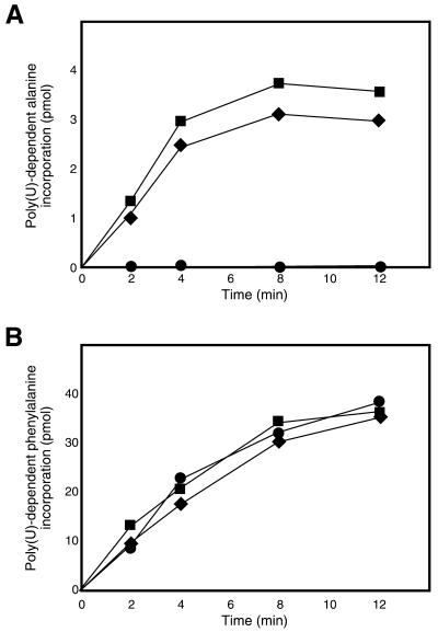
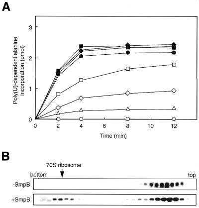
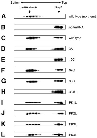
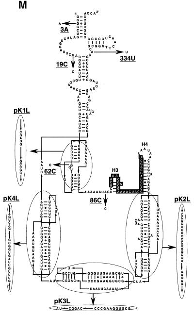
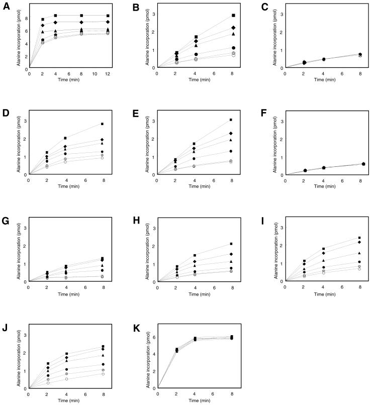
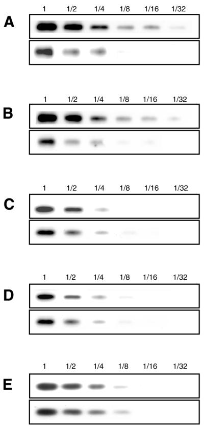
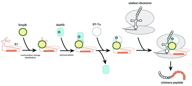
tmRNA has a dual function as a tRNA and an mRNA to facilitate trans-translation, in which a ribosome can switch between translation of a truncated mRNA and the tmRNA's tag sequence. SmpB is a tmRNA binding protein that has been identified to be essential for trans-translation in vivo. To further study the function of SmpB, an S30 fraction from an Escherichia coli strain, in which the set of genes for SmpB and tmRNA has been deleted from the genome, and His-tagged SmpB active in trans-translation were prepared. The SmpB-depleted S30 fraction had an ability to facilitate poly(U)-dependent tag-peptide synthesis in vitro when purified His-tagged SmpB was exogenously added together with tmRNA, although SmpB was not required for in vitro poly(U)-dependent poly(Phe) synthesis. It was also found that depletion of SmpB leads to a decrease in the level of tmRNA in the cell. In addition, SmpB considerably enhanced the aminoacylation of tmRNA by alanyl-tRNA synthetase in vitro. The aminoacylation enhancement by SmpB, the binding of SmpB to tmRNA and the effect of depletion of SmpB on the expression level of tmRNA in the cell were all affected by some mutations in the tRNA-like domain which cause a defect in ribosome binding leading to a trans-translation deficiency. These results demonstrate that, via binding to the tRNA-like domain of tmRNA, SmpB plays various roles: rescuing the tmRNA molecule from degradation in the cell, enhancing the aminoacylation of tmRNA and mediating the binding of tmRNA to ribosome.


Figures

Figure 1
Figure 1
(A) Poly(U)-dependent alanine incorporation using the S30 fractions from ΔsmpBΔssrA cells with (squares) and without (circles) plasmid-encoded SmpB and from ΔssrA cells (diamonds) in the presence of exogenous tmRNA. (B) Poly(U)-dependent phenylalanine incorporation using the S30 fractions from ΔsmpBΔssrA cells with (squares) and without (circles) plasmid-encoded SmpB and from ΔssrA cells (diamonds) in the presence of exogenous tmRNA.
Figure 2
Figure 2
(A) The effect of the addition of purified His-tagged SmpB into the S30 fractions from ΔsmpBΔssrA cells on poly(U)-dependent alanine incorporation in vitro. 0 (open circles), 0.05 (open triangles), 0.1 (open diamonds), 0.2 (open squares), 0.4 (closed circles), 0.8 (closed triangles), 1.6 (closed diamonds) and 3.2 µM (closed squares) of His-tagged SmpB were added to the reaction. (B) The effect of purified His-tagged SmpB on the ribosome binding of tmRNA in vitro.
Figure 3
Figure 3
Interaction of SmpB with tmRNA variants. (A) tmRNA fractionated by centrifugation on a glycerol density gradient was detected by northern hybridization. His-tagged SmpB alone (B) or mixed with wild-type tmRNA (C), 3A (D), 19C (E), 62C (F) 86C (G), 334U (H), pK1L (I), pK2L (J), pK3L (K) or pK4L (L) was fractionated by glycerol density gradient centrifugation and was detected by western blotting using an antibody raised against SmpB. (M) Mutations designated on a secondary structure model of E.coli tmRNA. The tag-encoded sequence highlighted by white with a black background is surrounded by four pseudoknots (PK1–PK4). Non-Watson–Crick base pairs are shown by open circles. This RNA has two tRNA-specific modified nucleotides, 5-methyl U and pseudouridine in the T-loop (6), indicated as T and Ψ, respectively. Arrows indicate the mutations used in this study.
Figure 3
Figure 3
Interaction of SmpB with tmRNA variants. (A) tmRNA fractionated by centrifugation on a glycerol density gradient was detected by northern hybridization. His-tagged SmpB alone (B) or mixed with wild-type tmRNA (C), 3A (D), 19C (E), 62C (F) 86C (G), 334U (H), pK1L (I), pK2L (J), pK3L (K) or pK4L (L) was fractionated by glycerol density gradient centrifugation and was detected by western blotting using an antibody raised against SmpB. (M) Mutations designated on a secondary structure model of E.coli tmRNA. The tag-encoded sequence highlighted by white with a black background is surrounded by four pseudoknots (PK1–PK4). Non-Watson–Crick base pairs are shown by open circles. This RNA has two tRNA-specific modified nucleotides, 5-methyl U and pseudouridine in the T-loop (6), indicated as T and Ψ, respectively. Arrows indicate the mutations used in this study.
Figure 4
Figure 4
The effect of SmpB on aminoacylation of tmRNA by 8.5 × 10⁻² U (A) and 8.5 × 10⁻³ U (B) of alanyl-tRNA synthetase. The effect of SmpB on aminoacylation of tmRNA mutants 19C (C), 62C (D) 86C (E), 334U (F), pK1L (G), pK2L (H), pK3L (I), pK4L (J) and E.coli tRNAAla (K), using 8.5 × 10⁻³ U of alanyl-tRNA synthetase. 0 µM (open circles), 0.1 µM (hatched circles), 0.5 µM (closed circles), 1 µM (closed triangles), 2 µM (closed diamonds) and 4 µM (closed squares) SmpB were added to the reaction of aminoacylation.
Figure 5
Figure 5
The expression levels of tmRNA in the cells of ΔssrA (top) and ΔsmpBΔssrA (bottom) strains carrying the low-copy number plasmid-encoded (A) wild-type tmRNA, (B) 3A, (C) 19C and (D) 334U mutants. (E) The expression levels of 5S ribosomal RNA in the cells of two strains carrying the low-copy number plasmid-encoded wild-type tmRNA. Varying dilutions of total RNA from the cell extract of each strain were electrophoresed on a 1.5% agarose gel containing 6.3% formaldehyde, and were then blotted onto a nylon membrane. tmRNA was detected by northern hybridization using a 3′-digoxygenin-labeled oligodeoxyribonucleotide. No degradation products of the tmRNA molecule were detected in the lower part of the gel irrespective of the presence or absence of SmpB. Presumably the primary degradation induces further degradation in the cell.
Figure 6
Figure 6
A possible pathway of the tmRNA complex formation for trans-translation. Judging from the dissociation constant with tmRNA of 10 nM or lower (36,44), the association of S1 might occur in the early stage of the complex formation. This model adopts a likely complex of SmpB, EF-Tu, S1 and tmRNA as a complex just prior to entrance to the stalled ribosome, as mentioned by Wower et al. (44). Some other possible factors such as phosphoribosyl pyrophosphate synthase, RNase R, YfbG (32) and tRNAAla (46) that have not yet been identified to be essential for trans-translation are not shown here.

References

    1. Williams K.P. (2000). The tmRNA website. Nucleic Acids Res., 28, 168. - PMC - PubMed
    1. Zwieb C. and Wower,J. (2000). (tmRDB) tmRNA database. Nucleic Acids Res., 28, 169–170. - PMC - PubMed
    1. Karzai A.W., Roche,E.D. and Sauer,R.T. (2000) The SsrA–SmpB system for protein tagging, directed degradation and ribosome rescue. NatureStruct. Biol., 7, 449–455. - PubMed
    1. Ushida C., Himeno,H., Watanabe,T. and Muto,A. (1994) tRNA-like structures in 10Sa RNAs of Mycoplasma capricolum and Bacillus subtilis. Nucleic Acids Res., 22, 3392–3396. - PMC - PubMed
    1. Komine Y., Kitabatake,M., Yokogawa,T., Nishikawa,K. and Inokuchi,H. (1994) A tRNA-like structure is present in 10Sa RNA, a small stable RNA from Escherichia coli. Proc. Natl Acad. Sci. USA, 91, 9223–9227. - PMC - PubMed

Publication types