. 2002 Apr 1;30(7):1695-703.
doi: 10.1093/nar/30.7.1695.

The terminase subunits pUL56 and pUL89 of human cytomegalovirus are DNA-metabolizing proteins with toroidal structure

Affiliations

The terminase subunits pUL56 and pUL89 of human cytomegalovirus are DNA-metabolizing proteins with toroidal structure

Hanno Scheffczik et al. Nucleic Acids Res. .

Abstract

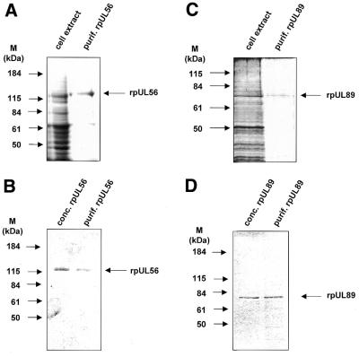
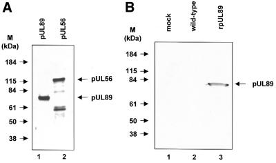
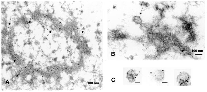
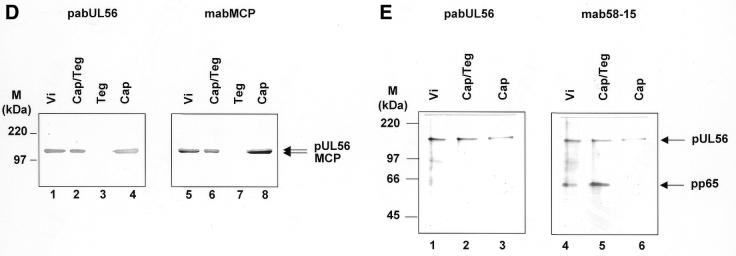
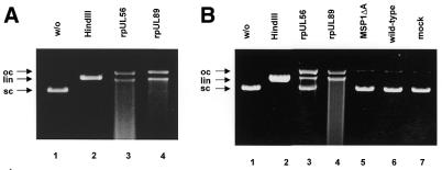
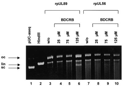
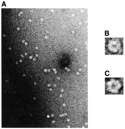
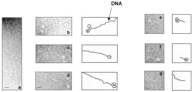
Herpesvirus DNA packaging involves binding and cleavage of DNA containing the specific DNA-packaging motifs. Here we report a first characterization of the terminase subunits pUL56 and pUL89 of human cytomegalovirus (HCMV). Both gene products were shown to have comparable nuclease activities in vitro. Under limiting protein concentrations the nuclease activity is enhanced by interaction of pUL56 and pUL89. High amounts of 2-bromo-5,6-dichloro-1-beta-D-ribofuranosyl benzimidazole partially inhibited the pUL89-associated nuclease activity. It was demonstrated that pUL56 is able to bind to nucleocapsids in vivo. Electron microscopy (EM) and image analysis of purified pUL56 revealed that the molecules occurred as a distinct ring-shaped structure with a pronounced cleft. EM analysis of purified pUL89 demonstrated that this protein is also a toroidal DNA-metabolizing protein. Upon interaction of pUL56 with linearized DNA, the DNA remains uncut while the cutting event itself is mediated by pUL89. Using biochemical assays in conjunction with EM pUL56 was shown to (i) bind to DNA and (ii) associate with the capsid. In contrast to this, EM analysis implied that pUL89 is required to effect DNA cleavage. The data provide the first insights into the terminase-dependent viral DNA-packaging mechanism of HCMV.


Figures

Figure 1
Figure 1
SDS–PAGE of cell lysates and purified proteins. (A) Aliquots of extracts from High Five cells infected with baculovirus UL56 (cell extract) and rpUL56 obtained after a two-step purification using anion exchange followed by gel permeation chromatography (purif. rpUL56) were subjected to SDS–PAGE prior to staining with silver. (B) Aliquots after purification (purif. rpUL56) and following spin concentration (conc. rpUl56) were subjected to SDS–PAGE prior to staining with Coomassie brilliant blue. (C) Aliquots of extracts from High Five cells infected with baculovirus UL89 (cell extract) and rpUL89 obtained after two-step purification using anion exchange followed by gel permeation chromatography (purif. rpUL89) were subjected to SDS–PAGE prior to staining with silver. (D) Aliquots after purification (purif. rpUL89) and following spin concentration (conc. rpUL89) were subjected to SDS–PAGE prior to staining with Coomassie brilliant blue. Molecular weight markers (M) are indicated on the left; the positions of the proteins are indicated on the right.
Figure 2
Figure 2
Identification of HCMV pUL89 in vitro translated or synthesized in insect cells infected with recombinant baculovirus UL89 or baculoviurs UL56. (A) Aliquots of pUL89 in vitro translation products were subjected to SDS–PAGE (8% polyacrylamide) prior to detection by autoradiography. (B) Aliquots of extracts from mock-transfected High Five cells (lane 1) or High Five cells infected with wild-type baculovirus (lane 2) or purified recombinant rpUL89 (lane 3) were separated by SDS–PAGE followed by immunoblotting with the specific anti-Xpress antibody against Xpress-tagged pUL89. Molecular weight standards (M) are indicated on the left; the positions of pUL89 and pUL56 are indicated by arrows.
Figure 3
Figure 3
Association of pUL56 with viral capsids. (A) Immunoelectron microscopy (IEM) analysis of an ultrathin section of HCMV-infected HFF cells. Immunogold labeling was carried out using pAbUL56 (dilution 1:10) and a goat anti-human antibody conjugated with colloidal gold (bead diameter 5 nm, dilution 1:50). (B) IEM analysis with double labeling was performed with (i) pAbUL56 and goat anti-human antibody conjugated with colloidal gold (bead diameter 10 nm) and (ii) mAb against pUL44 (dilution 1:10) and a goat anti-mouse antibody conjugated with colloidal gold (bead diameter 5 nm). (C) Immunogold labeling of purified capsids. Capsids were labeled with pAbUL56 and protein A conjugated with 5 nm colloidal gold. Arrows indicated capsids. The scale bars represent 100 nm. (D and E) Extracellular virions (vi) were separated into nucleocapsid/tegument (Cap/Teg), tegument (Teg) and nucleocapsid (Cap) components and analyzed by immunoblotting with pAbUL56 and an antibody against MCP (D) or with pAbUL56 and an antibody against pp65 (E). The arrows indicate the positions of pUL56, MCP and pp65 and the molecular weight standards (M) are indicated on the left.
Figure 3
Figure 3
Association of pUL56 with viral capsids. (A) Immunoelectron microscopy (IEM) analysis of an ultrathin section of HCMV-infected HFF cells. Immunogold labeling was carried out using pAbUL56 (dilution 1:10) and a goat anti-human antibody conjugated with colloidal gold (bead diameter 5 nm, dilution 1:50). (B) IEM analysis with double labeling was performed with (i) pAbUL56 and goat anti-human antibody conjugated with colloidal gold (bead diameter 10 nm) and (ii) mAb against pUL44 (dilution 1:10) and a goat anti-mouse antibody conjugated with colloidal gold (bead diameter 5 nm). (C) Immunogold labeling of purified capsids. Capsids were labeled with pAbUL56 and protein A conjugated with 5 nm colloidal gold. Arrows indicated capsids. The scale bars represent 100 nm. (D and E) Extracellular virions (vi) were separated into nucleocapsid/tegument (Cap/Teg), tegument (Teg) and nucleocapsid (Cap) components and analyzed by immunoblotting with pAbUL56 and an antibody against MCP (D) or with pAbUL56 and an antibody against pp65 (E). The arrows indicate the positions of pUL56, MCP and pp65 and the molecular weight standards (M) are indicated on the left.
Figure 4
Figure 4
Nuclease activity of HCMV rpUL56 and rpUL89. (A) Nuclease assay with purified proteins. Lane 1, pUC-aseq; lane 2, incubation in the presence of HindIII; lane 3, pUC-aseq incubated with purified rpUL56; lane 4, pUC-aseq incubated with purified rpUL89. (B) Nuclease assay with pBR322 as substrate. Lane 1, plasmid pBR322 in the absence of protein; lane 2, pBR322 with the restriction endonuclease HindIII; lane 3, pBR322 incubated with purified rpUL56; lane 4, pBR322 incubated with purified rpUL89; lane 5, incubation with purified MSP1ΔA; lane 6, pBR322 incubated with wild-type baculovirus-infected extracts; lane 7, pBR322 incubated in the presence of mock-infected extracts. The arrows indicate three different plasmid DNA forms: open circular molecules (oc), linear forms (lin) and supercoiled DNA (sc).
Figure 5
Figure 5
Influence of BDCRB on nuclease activity. Lane 1, pUC-aseq alone; lane 2, pUC-aseq treated with HindIII; lane 3, pUC-aseq plus purified rpUL56; lane 4, pUC-aseq incubated with rpUL89 and 25 µM BDCRB; lane 5, pUC-aseq incubated with rpUL89 and 75 µM BDCRB; lane 6, pUC-aseq incubated with rpUL89 and 125 µM BDCRB; lane 7, incubation with rpUL56; lane 8, incubation with rpUL56 and 25 µm BDCRB; lane 9, incubation with rpUL56 and 75 µM BDCRB; lane 10, incubation with rpUL56 and 125 µM BDCRB. The arrows indicate three different plasmid forms: open circular (oc), linear (lin) and supercoiled molecules (sc).
Figure 6
Figure 6
Nuclease activity with limited amounts of rpUL56 and rpUL89. Nuclease assay with purified proteins. Lane 1, plasmid pUC-aseq in the absence of protein; lane 2, pUC-aseq treated with the restriction endonuclease HindIII; lane 3, pUC-aseq incubated with rpUL56 (1.2 µg/ml); lane 4, pUC-aseq incubated with rpUL89 (1.2 µg/ml); lane 5, pUC-aseq incubated with rpUL56 and rpUL89 (1.2 µg/ml each). The arrows indicate three different plasmid DNA forms: open circular molecules (oc), linear forms (lin) and supercoiled DNA (sc).
Figure 7
Figure 7
Electron microscopic analysis of purified rpUL56 after negative staining. (A) An overview. (B and C) Representative molecules belonging to the two different projections (views 1 and 2, respectively). Note the toroidal structure of both projections and the pronounced cleft at the 9 o’clock position in (C). Averaged projections after digital image analysis are depicted in (D) (view 1) and (E) (view 2). The scale bars correspond to 50 (A), 10 (B and C) and 5 nm (D and E).
Figure 8
Figure 8
Electron microscopy of purified rpUL89. (A) An overview of rpUL89 negative stained with uranyl acetate. (B and C) Representative molecules. The scale bars correspond to 50 (A) and 1 nm (B and C).
Figure 9
Figure 9
Representative electron micrographic projections of linearized pUC-aseq after incubation with rpUL56 (ad) and rpUL56 + rpUL89 (eg). The sections show an ∼1 µM DNA molecule. The scale bars correspond to 10 nm.
Figure 10
Figure 10
Preliminary, schematic model of HCMV pUL56 depicting the proposed silent (A) and active (B) states. In the active state, DNA is thought to be encased by the protein. The proposed conformational changes might be a prerequisite for DNA binding of pUL56. HCMV pUL56 drawn to show the different forms as top and bottom views of pUL56 (C and D).

References

    1. Chou J. and Roizman,B. (1985) The isomerization of the herpes simplex virus 1 genome: identification of the cis-acting and recombination sites within the domain of the a sequence. Cell, 41, 803–811. - PubMed
    1. Deiss L.P., Chou,J. and Frenkel,N. (1986) Functional domains within the a sequence involved in the cleavage-packaging of herpes simplex virus DNA. J. Virol., 59, 605–618. - PMC - PubMed
    1. Spaete R.R. and Mocarski,E.S. (1985) The a sequence of the cytomegalovirus genome functions as a cleavage/packaging signal for herpes simplex virus defective genomes. J. Virol., 54, 817–824. - PMC - PubMed
    1. Black L.W. (1988) DNA packaging in dsDNA bacteriophages. In Calendar,R. (ed.), The Bacteriophages. Plenum, New York, NY, pp. 321–373.
    1. Murialdo H. and Becker,A. (1978) Head morphogenesis of complex double-stranded deoxyribonucleic acid bacteriophages. Microbiol. Rev., 42, 529–576. - PMC - PubMed

Publication types

MeSH terms