. 2002 Apr 2;99(7):4391-6.
doi: 10.1073/pnas.032074999. Epub 2002 Mar 26.

Endothelial cells derived from human embryonic stem cells

Affiliations

Endothelial cells derived from human embryonic stem cells

Shulamit Levenberg et al. Proc Natl Acad Sci U S A. .

Abstract

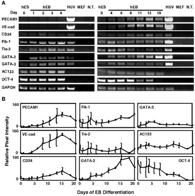
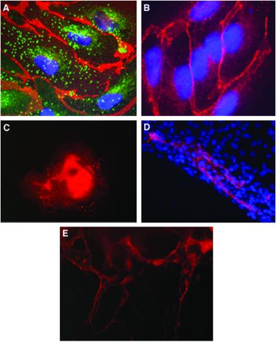
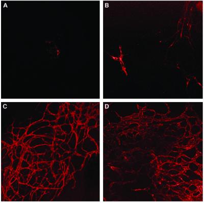
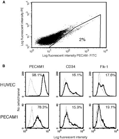
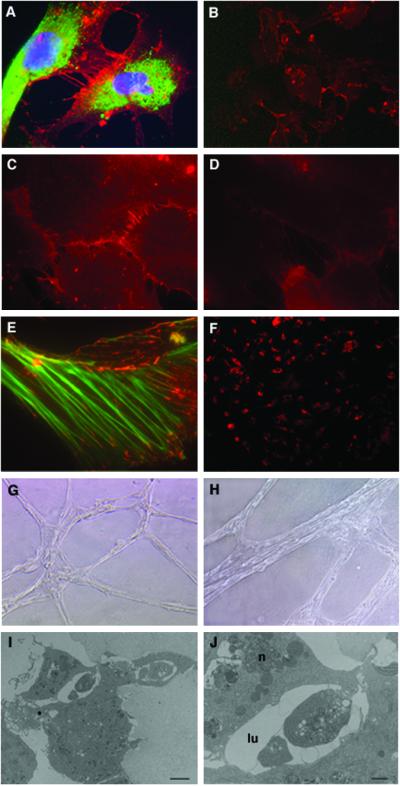
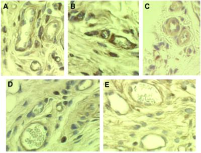
Human embryonic stem cells have the potential to differentiate into various cell types and, thus, may be useful as a source of cells for transplantation or tissue engineering. We describe here the differentiation steps of human embryonic stem cells into endothelial cells forming vascular-like structures. The human embryonic-derived endothelial cells were isolated by using platelet endothelial cell-adhesion molecule-1 (PECAM1) antibodies, their behavior was characterized in vitro and in vivo, and their potential in tissue engineering was examined. We show that the isolated embryonic PECAM1+ cells, grown in culture, display characteristics similar to vessel endothelium. The cells express endothelial cell markers in a pattern similar to human umbilical vein endothelial cells, their junctions are correctly organized, and they have high metabolism of acetylated low-density lipoprotein. In addition, the cells are able to differentiate and form tube-like structures when cultured on matrigel. In vivo, when transplanted into SCID mice, the cells appeared to form microvessels containing mouse blood cells. With further studies, these cells could provide a source of human endothelial cells that could be beneficial for potential applications such as engineering new blood vessels, endothelial cell transplantation into the heart for myocardial regeneration, and induction of angiogenesis for treatment of regional ischemia.


Figures

Figure 1
Figure 1
Endothelial gene expression in hES-derived EBs by RT-PCR analysis. (A) RNA was isolated from undifferentiated hES cells and from hEBs at different time points (days) during differentiation and subjected to RT-PCR analysis. The negative controls, no template (N.T.) and MEF, and the HUVEC positive control (HUV) are shown to the right. (B) Quantitative analysis of gene expression. Relative pixel intensity corresponds to gene expression level; for each time point, mean pixel intensities of each band were measured and normalized to mean pixel intensities of GAPDH band. The results shown are mean values ±SD of three different experiments.
Figure 2
Figure 2
Expression of endothelial cell markers in vessel-like structure within hEBs. (A) EBs at day 13 stained with human PECAM1 antibodies (red), vWF antibodies (green), and DAPI for nuclear staining (blue). PECAM1 is organized at cell–cell junctions, whereas vWF is found in organelles in the cytoplasm. (B) EB cells stained with human VE-cadherin antibodies (red) and DAPI (blue) (magnification, ×1,000). (C) Low magnification (×100) of EB stained with PECAM1 antibodies. (D) Areas of PECAM1+ cells (red) within part of an EB, organized in elongated clusters. Cell nuclei stained with DAPI (blue) (magnification, ×400). (E) Channels forming PECAM1+ cells within a 13-day-old EB (magnification, ×200).
Figure 3
Figure 3
Confocal microscopy of EBs stained for PECAM1 showing three-dimensional network formations, vascular-like channels. (A) 4-day-old EB. (B) 6-day-old EB. (C) 10-day-old EB. (D) 13-day-old EB. Notice the intensive and complicated vascular network developed at day 10 of 13-day-old EB. (magnification, ×100).
Figure 4
Figure 4
Isolation of endothelial cells from human embryoid bodies by using fluorescent-labeled anti-PECAM1 antibodies and analysis of the sorted cells. (A) EBs at day 13 were dissociated and incubated with PECAM1 antibodies. Fluorescent-labeled cells were isolated by using a flow cytometry cell sorter. (B) Flow cytometric analysis of endothelial cell markers in PECAM1+ cells grown in culture for six passages and HUVEC cells. The cells were dissociated and incubated with either isotype control (dashed lines) or antigen-specific antibodies, as indicated (solid lines). Percent positive cells are shown.
Figure 5
Figure 5
Characterization of hES-derived endothelial cells grown in culture. (A) Immunofluorescence staining of PECAM1 (red) at cell–cell junctions and vWF (green) in the cytoplasm. The nuclei are stained with DAPI (blue). (B) Cells stained for PECAM1. (C) N-cadherin and (D) VE-cadherin staining in cell–cell adherent junctions. (E) Double staining for vinculin (red) and actin (green). Vinculin is found in both focal contacts and cell–cell adherent junctions where it associates with actin stress fiber ends (magnification: A and CE, ×1,000; B, ×200). (F) Uptake of Dill-labeled ac-LDL by PECAM1+ cells. (G and H) Cords formation by PECAM1+ cells 24 h (G) or 3 days (H) after seeding the cells in matrigel (magnification: G, ×100; H, ×200). (I) Electron microscopy of the cord cross-section showing lumen formation (bar = 2 μm) and (J) higher magnification of the lumen (lu) area showing cell–cell interactions closing the lumen and the nucleus (n) of one cell (bar = 8 μm).
Figure 6
Figure 6
Transplantation of embryonic endothelial cells (PECAM1+) in SCID mice. PECAM1+ cells were seeded onto PLLA/PLGA polymer scaffolds as described in Materials and Methods. The cells plus scaffolds were implanted s.c. in the dorsal region of 4-week-old SCID mice. (AC) Immunoperoxidase (brown) staining of 7-day implants with anti-human PECAM1 antibodies and (D and E) of 14-day implants with anti-human CD34 antibodies, showing microvessels that are immunoreactive with these human-specific antibodies. Some of these human-positive microvessels have mouse blood cells in their lumen (magnification, ×400).

References

    1. Niklason L E, Gao J, Abbott W M, Hirschi K K, Houser S, Marini R, Langer R. Science. 1999;284:489–493. - PubMed
    1. Kawamoto A, Gwon H C, Iwaguro H, Yamaguchi J I, Uchida S, Masuda H, Silver M, Ma H, Kearney M, Isner J M, Asahara T. Circulation. 2001;103:634–637. - PubMed
    1. Vittet D, Prandini M H, Berthier R, Schweitzer A, Martin-Sisteron H, Uzan G, Dejana E. Blood. 1996;88:3424–3431. - PubMed
    1. Yancopoulos G D, Klagsbrun M, Folkman J. Cell. 1998;93:661–664. - PubMed
    1. Yamashita J, Itoh H, Hirashima M, Ogawa M, Nishikawa S, Yurugi T, Naito M, Nakao K. Nature (London) 2000;408:92–96. - PubMed

Publication types

MeSH terms